Fraud Blocker Skip to main content

Spinal Collection

"Unlocking the Secrets of Spinal Harmony: Exploring Chakras and the Nervous System" Delve into the intricate world of the spinal column

Background imageSpinal Collection: Chakras and Nervous System

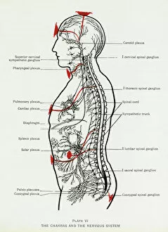
Chakras and Nervous System
How the chakras interact with the nervous system. Date: 1927

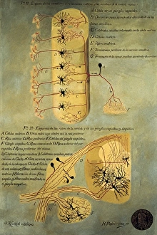
Background imageSpinal Collection: RAMON Y CAJAL, Santiago (1852-1934). Spanish

RAMON Y CAJAL, Santiago (1852-1934). Spanish doctor and histologist, Nobel Prize in 1906. Histological schema of the spinal cord. Original drawing by Ram󮠹 Cajal. SPAIN. Madrid

Background imageSpinal Collection: Diagram of the human brain and spinal column

Diagram of the human brain and spinal column
A diagram of the brain and spinal column, including the vertebrae

Background imageSpinal Collection: Calots spinal surgery, 19th century

Calots spinal surgery, 19th century
Calots spinal surgery, 19th-century artwork. This operation is being carried out by the French surgeon Jean-Francois Calot (1861-1944) on a condition known as Potts disease

Background imageSpinal Collection: Upper body skeleton, computer artwork

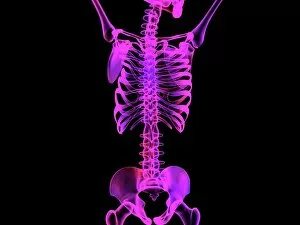
Upper body skeleton, computer artwork
Upper body skeleton. Computer artwork of a human skeleton showing the bones of the upper body. At the top is the base of the skull and the jaw bone
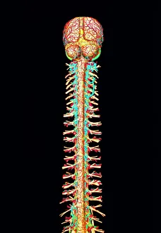
Background imageSpinal Collection: Illustration of the human spinal cord and brain

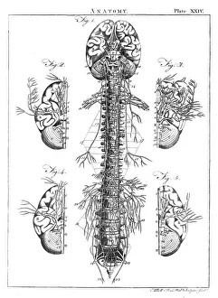
Illustration of the human spinal cord and brain
Spinal cord. Computer-enhanced historical illustr- ation of the spinal cord and brain of a human. Both organs are seen without the skeletal structures (such as the skull & vertebrae)

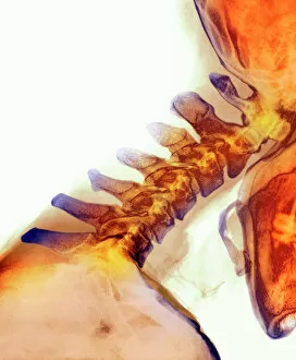
Background imageSpinal Collection: Neck vertebrae extended, X-ray

Neck vertebrae extended, X-ray
Bending of the neck. Coloured X-ray of a side view of the neck of a man showing extension of healthy cervical vertebrae (bones)

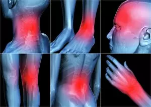
Background imageSpinal Collection: Body pain, artwork

Body pain, artwork
Body pain. Computer artwork showing pain (represented by the red areas) in six different body areas. From top left to lower right, the body areas are: the neck, ankle, head, knee, lower back and wrist

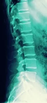
Background imageSpinal Collection: Normal spine, X-ray

Normal spine, X-ray
Normal spine. Coloured profile X-ray showing bones of the spine (vertebrae, centre). The front of the body is at right. The ribs (upper right) and the pelvis (bottom) are also seen

Background imageSpinal Collection: Normal neck, X-ray

Normal neck, X-ray
Noraml neck. X-ray of the healthy cervical (neck) spine of a 29 year old male patient

Background imageSpinal Collection: Lower back pain, conceptual artwork

Lower back pain, conceptual artwork
Lower back pain. Conceptual computer artwork showing pain (represented as the red area) in the lower back. The bones (white) of the lower torso and arms are shown within an outline (blue)

Background imageSpinal Collection: Upper back pain, conceptual artwork

Upper back pain, conceptual artwork
Upper back pain. Conceptual computer artwork showing pain (represented as the red area) in the upper back. The bones (white) of the torso and arms are shown within an outline (blue)

Background imageSpinal Collection: Nervous System 18th C

Nervous System 18th C
The nervous system of the spine and the front body according to Vieuffens Date: Circa 1760

Background imageSpinal Collection: Woman stretching

Woman stretching
MODEL RELEASED. Woman stretching

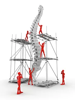
Background imageSpinal Collection: Spine with workers, spine repair F007 / 9907

Spine with workers, spine repair F007 / 9907
Conceptual computer artwork depicting a spine being repaired

Background imageSpinal Collection: Slipped intervertebral disc, artwork

Slipped intervertebral disc, artwork
Slipped intervertebral disc, computer artwork. Intervertebral discs (red) are pads of cartilage between the bones (vertebrae) of the spine

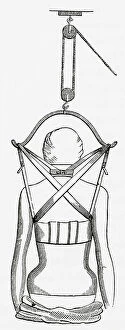
Background imageSpinal Collection: Suspension apparatus for use in spinal disease. From The Household Physician

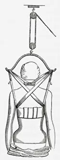
Suspension apparatus for use in spinal disease. From The Household Physician, published c.1898 (engraving)
6116635 Suspension apparatus for use in spinal disease. From The Household Physician, published c.1898 (engraving); Photo © Ken Welsh. All rights reserved 2024.

Background imageSpinal Collection: Human anatomy, nervous system, brain and spinal cord

Human anatomy, nervous system, brain and spinal cord. Handcolored engraving by Fournier after an illustration by Martin St

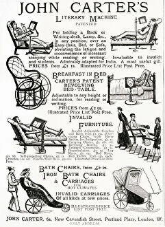
Background imageSpinal Collection: Advert for John Carters invalid chairs 1884

Advert for John Carters invalid chairs 1884
Selection of invalid and students chairs, including holding a book or writing at a desk, lamp in any position over an easy chair

Background imageSpinal Collection: California Usa America American Death Valley

California Usa America American Death Valley
california, usa, america, american, death, valley, abandoned, cabin, shed, wood, wooden, makeshift, building, buildings, ramshackle, animal, skulls, skull, bone, bones, sand, springs, north, end

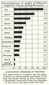
Background imageSpinal Collection: Starvation, degrees of wasting (litho)

Starvation, degrees of wasting (litho)
5998374 Starvation, degrees of wasting (litho) by English School, (20th century); Private Collection; (add.info.: Starvation, degrees of wasting)

Background imageSpinal Collection: Asian tapeworm and sheep tapeworm larva

Asian tapeworm and sheep tapeworm larva
Asian tapeworm, Bothriocephalus acheilognathi (Botryocephalus latus), freshwater fish parasite, and Coenurus cerebralis, larva of the tapeworm Multiceps multiceps, found in sheeps brains

Background imageSpinal Collection: Advert for Carters spinal carriage 1906

Advert for Carters spinal carriage 1906
Carters bath chair and spinal carriage in wood and wicker-work Date: 1906

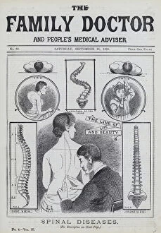
Background imageSpinal Collection: Spinal diseases (engraving)

Spinal diseases (engraving)
5223909 Spinal diseases (engraving) by English School, (19th century); Private Collection; (add.info.: Spinal diseases. Illustration for The Family Doctor and Peoples Medical Adviser)

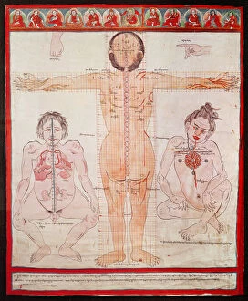
Background imageSpinal Collection: Anatomical Chart, 1938 (pen and watercolour on cloth laid on canvas)

Anatomical Chart, 1938 (pen and watercolour on cloth laid on canvas)
CHT163244 Anatomical Chart, 1938 (pen and watercolour on cloth laid on canvas) by Tibetan School; Wellcome Museum, London

Background imageSpinal Collection: Poor posture for reading and writing (engraving)

Poor posture for reading and writing (engraving)
3097123 Poor posture for reading and writing (engraving) by German School, (19th century); Private Collection; (add.info.: Poor posture for reading and writing)

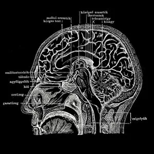
Background imageSpinal Collection: Human head section

Human head section
Longitudinal section of the human head

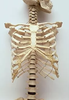
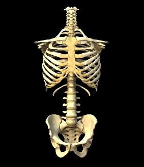
Background imageSpinal Collection: Human rib cage, jaw bones, neck vertabrae leading to sternum, collar bones and ribcage

Human rib cage, jaw bones, neck vertabrae leading to sternum, collar bones and ribcage, spinal column continuing to pelvis, front view

Background imageSpinal Collection: Advert for Carter wounded or invalid chairs 1896

Advert for Carter wounded or invalid chairs 1896
For holding a book or writing at a desk in any position over an easy chair, bed or sofa obviating fatigue and stooping. In valuable to invalids and students. 1896

Background imageSpinal Collection: Medicine. Study of anatomy by Leonardo Da Vinci. 15th centur

Medicine. Study of anatomy by Leonardo Da Vinci. 15th centur
Study of anatomy by Leonardo Da Vinci. 15th century. Skeletal structure. National Museum of Science and Technology Leonardo da Vinci. Milan. Italy

Background imageSpinal Collection: Human skeleton anatomy, artwork

Human skeleton anatomy, artwork
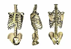
Human skeleton anatomy, computer artwork. Three views of the bones of the human torso, seen from an oblique frontal view (left), from the side (centre) and from the front (right)

Background imageSpinal Collection: Coccyx pain, conceptual artwork

Coccyx pain, conceptual artwork
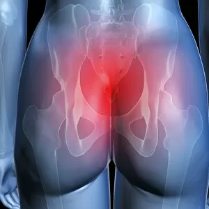
Coccyx pain. Conceptual computer artwork showing pain (represented as the red area) in the coccyx, the base of the spine. The bones (white) of the upper legs

Background imageSpinal Collection: Suspension apparatus for use in spinal disease. From The Household Physician, published c. 1898

Suspension apparatus for use in spinal disease. From The Household Physician, published c. 1898
Suspension apparatus for use in spinal disease. From The Household Physician, published c.1898

Background imageSpinal Collection: Lower half of human skeleton, jaw, spinal column, rib cage, pelvis, femurs, arms

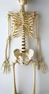
Lower half of human skeleton, jaw, spinal column, rib cage, pelvis, femurs, arms, hands and fingers, front view

Background imageSpinal Collection: android, bald, bikini, cloud, color image, concept, copy space, cyborg, day, digital composite

android, bald, bikini, cloud, color image, concept, copy space, cyborg, day, digital composite
Android women kneeling and laying on floor

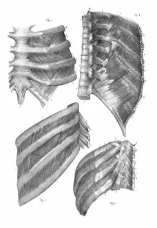
Background imageSpinal Collection: Intercostal nerves anatomy engraving 1866

Intercostal nerves anatomy engraving 1866
Atlas d anatomie descriptive du corps humain C. Bonamy - Paul Broca Victor Masson et Fils Paris 1866

Background imageSpinal Collection: Pelvic-femoral muscles, 1831 artwork

Pelvic-femoral muscles, 1831 artwork
Pelvic-femoral muscles, superficial layer. Part of the spine at top left. This anatomical artwork is plate 124 from volume 2 (1831) of Traite complet de l anatomie de l homme (1831-1854)

Background imageSpinal Collection: Hip joint bones and anatomy, artwork C014 / 2032

Hip joint bones and anatomy, artwork C014 / 2032
Hip joint bones and anatomy. Artwork of the bones that form the two hip joints (right and left). The hip is a ball-and-socket joint that provides the mobility needed for walking and running

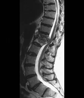
Background imageSpinal Collection: Tuberculosis of the spine, MRI scan

Tuberculosis of the spine, MRI scan
Tuberculosis of the spine. Magnetic resonance imaging (MRI) scan of a sagittal section through the spine of a 74-year-old patient with tuberculosis (TB) of the spine, or Potts disease

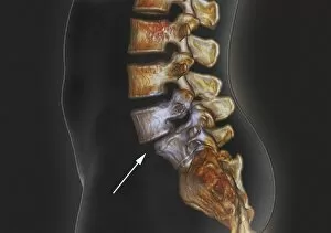
Background imageSpinal Collection: Spondylolisthesis, 3D CT scan

Spondylolisthesis, 3D CT scan
Spondylolisthesis. Coloured 3D computed tomography (CT) scan of a section through the lumbar (lower back) spine of a 39-year-old patient with spondylolisthesis

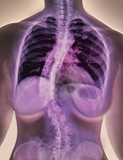
Background imageSpinal Collection: Scoliosis of the spine, X-ray C016 / 6566

Scoliosis of the spine, X-ray C016 / 6566
Scoliosis of the spine. Coloured X-ray of the chest of a 34 year old female patient with scoliosis (sideways curvature) of the spine

Background imageSpinal Collection: Australopithecus africanus pelvis, STS-14 C015 / 6919

Australopithecus africanus pelvis, STS-14 C015 / 6919
Australopithecus africanus pelvis (STS-14). This fossil specimen was discovered in 1947, in Sterkfontein, South Africa. The entire specimen consists of the pelvis, part of the vertebral column

Background imageSpinal Collection: Coccyx pain, artwork

Coccyx pain, artwork
Coccyx (tailbone) pain, computer artwork

Background imageSpinal Collection: Male torso skeleton

Male torso skeleton. The bones of the chest and abdomen surround and protect the organs and provide support for the body. The ribcage (upper frame) encloses the heart and lungs

Background imageSpinal Collection: Skeleton lifting a box incorrectly

Skeleton lifting a box incorrectly. Side view X- ray. This image shows the incorrect method of lifting heavy objects using the lower back muscles with the spine bent

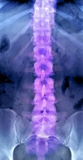
Background imageSpinal Collection: Normal lumbar spine, X-ray

Normal lumbar spine, X-ray
Normal lumbar spine. Coloured frontal X-ray showing bones of the lower (lumbar) spine (centre, purple). The bones of the spine (block-shaped) are called vertebrae

Background imageSpinal Collection: Bones of the human skeleton

Bones of the human skeleton
Bones of the skeleton. Artwork from Juan Valverde de Amuscos textbook Anatomia del Corpo Humano (Anatomy of the Human Body), published 1560. Fig. I is a view of the spine, from the left side. Figs

Background imageSpinal Collection: Potts disease in Egyptian tomb drawing

Potts disease in Egyptian tomb drawing
Tracing from an Egyptian tomb of a man suffering from Potts disease, or tuberculosis of the spine. The deformity is due to a bacterial infection in childhood which affects the front of the spinal


All Professionally Made to Order for Quick Shipping

"Unlocking the Secrets of Spinal Harmony: Exploring Chakras and the Nervous System" Delve into the intricate world of the spinal column, where ancient wisdom meets modern science. Discover how this remarkable structure, revered by pioneers like Ramon y Cajal, holds the key to our physical and spiritual well-being. Embark on a visual journey through time as you explore a diagram showcasing the human brain and spinal cord. Witness how these interconnected powerhouses orchestrate every aspect of our existence, from bodily functions to emotional experiences. Step back in time with Calots spinal surgery, an innovation from the 19th century that revolutionized medical practices. Marvel at illustrations depicting this groundbreaking procedure that aimed to alleviate pain and restore mobility. Peer into X-ray images capturing both normal necks and spines, revealing their intricate beauty hidden beneath our skin. Observe an extended neck vertebrae X-ray, shedding light on potential issues caused by misalignment or injury. Art comes alive as body pain is transformed into captivating artwork – a poignant reminder of the challenges faced by those afflicted with discomfort along their spine. Let your imagination soar as upper body skeletons take form through computer-generated artistry. Witness how lower back pain manifests itself in conceptual artwork – a vivid representation of its impact on daily life. Explore similar expressions for upper back pain; each stroke conveying tales of struggle yet resilience against this common ailment. Intriguingly juxtaposing these artistic interpretations are X-rays showcasing normal necks and spines - silent testaments to health and balance within our bodies' core structures. Uncover new perspectives on spinal health as you immerse yourself in this captivating collection.