
Slipped Disc Collection
"Exploring the Depths of Body Pain through Artwork: A Visual Journey into Slipped Discs" Lower back pain can be a debilitating experience


All Professionally Made to Order for Quick Shipping
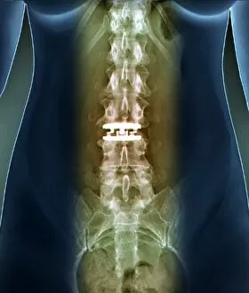
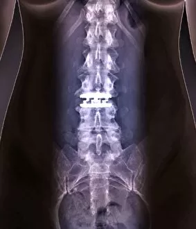
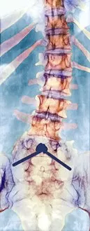
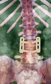
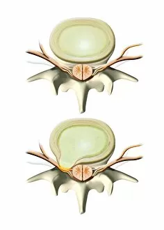
"Exploring the Depths of Body Pain through Artwork: A Visual Journey into Slipped Discs" Lower back pain can be a debilitating experience, but what if we could capture its essence through conceptual artwork? Dive into the depths of discomfort with this thought-provoking piece. Unveiling the unseen agony of upper back pain, this conceptual artwork delves into the complexities and struggles faced by those suffering from slipped discs. Prepare to have your perspective shifted. The intricate beauty of a slipped disc is unveiled in this captivating artwork, showcasing the delicate interplay between vertebrae and nerves that can cause immense pain and discomfort. Peering beneath the surface, an X-ray reveals pinned vertebrae caused by a slipped intervertebral disc—a visual representation that highlights both the fragility and resilience of our spinal structure. Neck pain takes on new meaning in this striking conceptual artwork, shedding light on its often overlooked impact on daily life. Let your imagination wander as you explore its hidden layers. Delving deeper into spinal health, an MRI scan unveils the aftermath of a spinal cord stroke—an eye-opening image that reminds us how vital it is to care for our backs and seek timely treatment when needed. Even our furry friends aren't spared from spine-related issues. An illustration showcases a prolapsed disc in a dog's spine—a reminder that these conditions can affect all creatures great and small. Witnessing sciatica's grip on one's body becomes possible with an MRI scan—revealing nerve compression like never before seen. Brace yourself for an enlightening journey through chronic lower back pain. Capturing both artistry and medical insight, witness lower back pain C016 / 9667 come alive through vivid imagery—an invitation to empathize with those who battle constant discomfort every day. Marvel at modern medicine's ingenuity as an artificial spinal disc takes center stage in an X-ray C016 / 6561.