Fraud Blocker Skip to main content

Sex Cell Collection

"Exploring the Intricate World of Sex Cells: A Journey through SEM Images" Delve into the mesmerizing realm of sex cells as we embark on a captivating journey

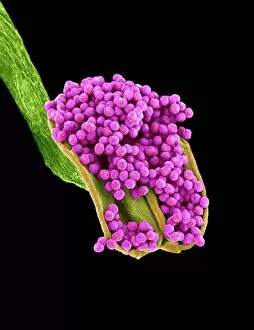
Background imageSex Cell Collection: Geranium anther, SEM

Geranium anther, SEM
Geranium anther. Coloured scanning electron micrograph (SEM) of pollen (pink) on the anther of a geranium flower (Geranium sp.)

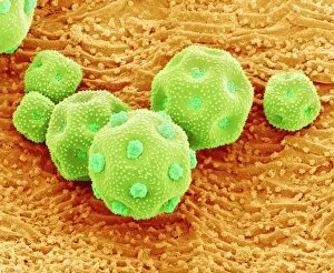
Background imageSex Cell Collection: Passion flower pollen, SEM

Passion flower pollen, SEM
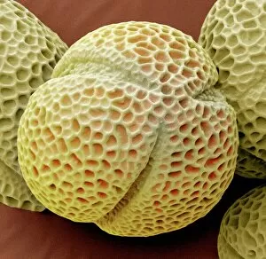
Passion flower pollen. Coloured scanning electron micrograph (SEM) of a passion flower (Passiflora caerulea) pollen grain. Pollen grains are the male gametes (sex cells) of a plant

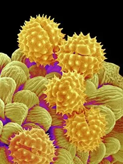
Background imageSex Cell Collection: Geranium pollen, SEM

Geranium pollen, SEM
Geranium pollen. Coloured scanning electron micrograph (SEM) of pollen (pink) on the stamen of a geranium flower (Geranium sp.)

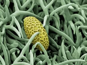
Background imageSex Cell Collection: Dandelion pollen grain, SEM

Dandelion pollen grain, SEM
Dandelion (Taraxacum officinale) pollen grain, coloured scanning electron micrograph (SEM). Pollen grains are reproductive structures produced by the male parts of flowering plants

Background imageSex Cell Collection: Pollen on a bee leg, SEM

Pollen on a bee leg, SEM
Pollen on a bee leg. Coloured scanning electron micrograph (SEM) of pollen grains (pale purple) on the leg of a bee (superfamily Apoidea). Pollen grains are the male sex cells of a flowering plant

Background imageSex Cell Collection: Philadelphia fleabane pollen grains, SEM

Philadelphia fleabane pollen grains, SEM
Philadelphia fleabane (Erigeron philadelphicus) pollen grains, coloured scanning electron micrograph (SEM). Pollen grains are reproductive structures produced by the male parts of flowering plants

Background imageSex Cell Collection: Gorse stigma with pollen grains, SEM

Gorse stigma with pollen grains, SEM
Gorse (Ulex europaeus) stigma (green) with pollen grains (yellow), coloured scanning electron micrograph (SEM). Pollen grains are reproductive structures produced by the male parts of flowering

Background imageSex Cell Collection: Lily pollen grain on rosemary leaf, SEM

Lily pollen grain on rosemary leaf, SEM
Lily pollen grain on rosemary leaf. Coloured scanning electron micrograph (SEM) of a pollen grain (yellow) from a lily flower (Lilium sp.)

Background imageSex Cell Collection: Forsythia pollen grains, SEM

Forsythia pollen grains, SEM
Forsythia pollen grains arranged in a spiral, coloured scanning electron micrograph (SEM). Pollen grains are reproductive structures produced by the male parts of flowering plants

Background imageSex Cell Collection: Chickweed pollen grains, SEM

Chickweed pollen grains, SEM
Chickweed pollen grains. Coloured scanning electron micrograph (SEM) of pollen grains (green) from a chickweed flower (Stellaria media)

Background imageSex Cell Collection: Hellebore pollen, SEM

Hellebore pollen, SEM
Hellebore (Helleborus sp.) pollen grains, coloured scanning electron micrograph (SEM). Magnification: x2000 when printed at 10 centimetres wide

Background imageSex Cell Collection: Germinating lily pollen, SEM

Germinating lily pollen, SEM
Germinating lily pollen. Coloured scanning electron micrograph (SEM) of lily (Lilium sp.) pollen grains that have sprouted pollen tubes. Pollen contains the male sex cells of a plant

Background imageSex Cell Collection: Pollination, SEM

Pollination, SEM
Pollination. Coloured scanning electron micrograph (SEM) of part of a flowers pistil with pollen grains attached. The pistil is the female reproductive part of a flower

Background imageSex Cell Collection: Deformed sperm cell, SEM

Deformed sperm cell, SEM
Deformed sperm cell. Coloured scanning electron micrograph (SEM) of double-tailed sperm cell, or spermatozoon. A sperm cells motility may be seriously affected by having multiple heavy tails

Background imageSex Cell Collection: Human genetic engineering

Human genetic engineering. Conceptual computer artwork representing the genetic engineering of humans. Sperm cells labelled with bar codes are seen

Background imageSex Cell Collection: Chickweed pollen grain, SEM

Chickweed pollen grain, SEM
Chickweed pollen grain. Coloured scanning electron micrograph (SEM) of a pollen grain (green) from a chickweed flower (Stellaria media)

Background imageSex Cell Collection: Basil pollen grain, SEM

Basil pollen grain, SEM
Basil pollen grain. Coloured scanning electron micrograph (SEM) of a pollen grain from the flower of a basil (Ocimum basilicum) plant. Pollen grains are the male gametes (sex cells) of a plant

Background imageSex Cell Collection: Fungal spores on pollen grain, SEM

Fungal spores on pollen grain, SEM
Fungal spores on pollen grain. Coloured scanning electron micrograph (SEM) showing fungal spores (round) on a pollen grain from a goji berry (wolfberry, Lycium sp.) plant

Background imageSex Cell Collection: Fertilisation, artwork F006 / 8739

Fertilisation, artwork F006 / 8739
Fertilisation, computer artwork

Background imageSex Cell Collection: Human sperm cells, artwork F006 / 8688

Human sperm cells, artwork F006 / 8688
Human sperm cells, computer artwork

Background imageSex Cell Collection: Ovulation, artwork C016 / 7537

Ovulation, artwork C016 / 7537
Ovulation, computer artwork

Background imageSex Cell Collection: Cowslip pollen grain, SEM C016 / 9031

Cowslip pollen grain, SEM C016 / 9031
Cowslip pollen grain. Coloured scanning electron micrograph (SEM) of pollen grain from a common cowslip (Primula veris) flower. Pollen grains are the male sex cells of a flowering plant

Background imageSex Cell Collection: Cowslip pollen grain, SEM C016 / 9030

Cowslip pollen grain, SEM C016 / 9030
Cowslip pollen grain. Coloured scanning electron micrograph (SEM) of pollen grain from a common cowslip (Primula veris) flower. Pollen grains are the male sex cells of a flowering plant

Background imageSex Cell Collection: Dwarf checkermallow pollen grain, SEM C016 / 9438

Dwarf checkermallow pollen grain, SEM C016 / 9438
Dwarf checkermallow pollen grain. Coloured scanning electron micrograph (SEM) of a pollen grain from a dwarf checkermallow (Sidalcea malviflora) flower

Background imageSex Cell Collection: Camellia pollen, SEM C015 / 9103

Camellia pollen, SEM C015 / 9103
Camellia pollen grains. Coloured scanning electron micrograph (SEM) of pollen grains from a Camellia sp. flower. The grains have a highly sculpted outer wall (exine)

Background imageSex Cell Collection: Geranium pollen, SEM C015 / 9155

Geranium pollen, SEM C015 / 9155
Geranium pollen. Coloured scanning electron micrograph (SEM) of pollen (pink) on the stamen of a geranium flower (Geranium sp.)

Background imageSex Cell Collection: Camellia pollen, SEM C015 / 9101

Camellia pollen, SEM C015 / 9101
Camellia pollen grains. Coloured scanning electron micrograph (SEM) of pollen grains from a Camellia sp. flower. The grains have a highly sculpted outer wall (exine)

Background imageSex Cell Collection: Camellia pollen, SEM C015 / 9089

Camellia pollen, SEM C015 / 9089
Camellia pollen grains. Coloured scanning electron micrograph (SEM) of pollen grains from a Camellia sp. flower. The grains have a highly sculpted outer wall (exine)

Background imageSex Cell Collection: Geranium pollen, SEM C015 / 9159

Geranium pollen, SEM C015 / 9159
Geranium pollen. Coloured scanning electron micrograph (SEM) of a pollen grain (pink) on the stamen of a geranium flower (Geranium sp.)

Background imageSex Cell Collection: Sperm cells, SEM C015 / 7842

Sperm cells, SEM C015 / 7842
Sperm cells. Coloured scanning electron micrograph (SEM) of sperm cells (spermatozoa). Each sperm cell consists of a head (round)

Background imageSex Cell Collection: Camellia pollen, SEM C015 / 9090

Camellia pollen, SEM C015 / 9090
Camellia pollen grains. Coloured scanning electron micrograph (SEM) of pollen grains from a Camellia sp. flower. The grains have a highly sculpted outer wall (exine)

Background imageSex Cell Collection: Geranium pollen, SEM C015 / 9156

Geranium pollen, SEM C015 / 9156
Geranium pollen. Coloured scanning electron micrograph (SEM) of pollen (pink) on the stamen of a geranium flower (Geranium sp.)

Background imageSex Cell Collection: Geranium pollen, SEM C015 / 9157

Geranium pollen, SEM C015 / 9157
Geranium pollen. Coloured scanning electron micrograph (SEM) of pollen (pink) on the stamen of a geranium flower (Geranium sp.)

Background imageSex Cell Collection: Geranium pollen, SEM C015 / 9158

Geranium pollen, SEM C015 / 9158
Geranium pollen. Coloured scanning electron micrograph (SEM) of a pollen grain (pink) on the stamen of a geranium flower (Geranium sp.)

Background imageSex Cell Collection: Sunflower pollen grain, SEM C018 / 0310

Sunflower pollen grain, SEM C018 / 0310
Sunflower pollen grain. Coloured scanning electron micrograph (SEM) of a pollen grain from the sunflower (Helianthus annuus)

Background imageSex Cell Collection: Sunflower pollen grains, SEM C018 / 0309

Sunflower pollen grains, SEM C018 / 0309
Sunflower pollen grains. Coloured scanning electron micrograph (SEM) of pollen grains from the sunflower (Helianthus annuus)

Background imageSex Cell Collection: Sperm cell, artwork C018 / 6996

Sperm cell, artwork C018 / 6996
Sperm cell anatomy. Cutaway computer artwork showing the internal structure of a sperm cell (spermatozoon), the male sex cell

Background imageSex Cell Collection: Sperm tails, TEM C014 / 1463

Sperm tails, TEM C014 / 1463
Sperm tails. Transmission electron micrograph (TEM) of sections through the tails of sperm (spermatozoa) cells at various levels

Background imageSex Cell Collection: Sperm tails, TEM C014 / 1465

Sperm tails, TEM C014 / 1465
Sperm tails. Transmission electron micrograph (TEM) of sections through the tails of sperm (spermatozoa) cells at various levels

Background imageSex Cell Collection: Sperm tails, TEM C014 / 1464

Sperm tails, TEM C014 / 1464
Sperm tails. Transmission electron micrograph (TEM) of sections through the tails of sperm (spermatozoa) cells at various levels

Background imageSex Cell Collection: Aubergine pollen grain, SEM

Aubergine pollen grain, SEM
Aubergine pollen grain. Coloured scanning electron micrograph (SEM) of a pollen grain from the flower of an aubergine (Solanum melongena, eggplant) plant

Background imageSex Cell Collection: Ovulation and fertilisation, artwork

Ovulation and fertilisation, artwork
Ovulation and fertilisation. Computer artwork of the female reproductive system, showing the development of an ovum (egg, small circle) inside an ovary (oval)

Background imageSex Cell Collection: Sperm cell anatomy, artwork C013 / 4648

Sperm cell anatomy, artwork C013 / 4648
Sperm cell anatomy. Cutaway computer artwork showing the internal structure of a sperm cell (spermatozoon), the male sex cell

Background imageSex Cell Collection: Scots pine pollen, SEM

Scots pine pollen, SEM
Scots pine pollen. Coloured scanning electron micrograph (SEM) of Scots pine (Pinus sylvestris) pollen grains. The pollen grains consist of a base (orange) with two air-filled

Background imageSex Cell Collection: Scots pine pollen grains, SEM

Scots pine pollen grains, SEM
Scots pine tree pollen grains. Coloured scanning electron micrograph (SEM) of Scots pine (Pinus sylvestris) pollen grains

Background imageSex Cell Collection: Liver flukes, transverse sections

Liver flukes, transverse sections
Liver flukes. Light micrographs of transverse sections through the male (top) and female (bottom) body areas of an adult liver fluke (Fasciola hepatica)

Background imageSex Cell Collection: Bee foot, SEM

Bee foot, SEM
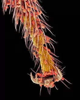
Bee foot. Coloured scanning electron micrograph (SEM) of the tip of a leg from a bee (superfamily Apoidea). The end of an insect leg consists of the final segment, which is called the tarsus

Background imageSex Cell Collection: Sperm in the testis, TEM

Sperm in the testis, TEM
Sperm cells. Coloured transmission electron micrograph (TEM) of a section through the testis, showing sperm cells. The heads of the sperm, which contain the genetic material, are red


All Professionally Made to Order for Quick Shipping

"Exploring the Intricate World of Sex Cells: A Journey through SEM Images" Delve into the mesmerizing realm of sex cells as we embark on a captivating journey, guided by high-resolution scanning electron microscope (SEM) images. Witness the intricate beauty and diversity that lies within these tiny reproductive units. Marvel at the Geranium anther, where delicate pollen grains are nestled in vibrant hues, ready to embark on their crucial mission of fertilization. The Passion flower pollen reveals its unique structure under SEM, showcasing its fascinating intricacies. Observe the Geranium pollen grain with its distinct features, highlighting nature's attention to detail. The Dandelion pollen grain captivates with its spherical shape and textured surface when examined closely under SEM. Discover how pollination occurs as you observe Pollen on a bee leg captured in stunning detail using SEM technology. Philadelphia fleabane pollen grains reveal themselves like miniature jewels upon closer inspection. Witness Gorse stigma adorned with numerous strikingly beautiful pollen grains through SEM imagery. A Lily pollen grain finds itself delicately perched upon a rosemary leaf, creating an enchanting contrast between textures and colors. The Forsythia pollen grains showcase their unique shapes and patterns when magnified using SEM techniques. Chickweed's microscopic yet elegant-looking pollen grains come alive in this visual exploration. Uncover the Hellebore's exquisite morphology through detailed SEM imaging—a testament to nature's artistry even at such minuscule scales. Finally, witness germinating lily pollen as it embarks on new life journeys under close scrutiny from our powerful microscope lenses. Embark on this captivating voyage into the world of sex cells—where beauty meets science—and marvel at nature's ingenuity revealed through astonishingly detailed SEM images capturing every intricate facet of these vital reproductive elements.