Fraud Blocker Skip to main content

Sectioned Collection (page 2)

"Exploring the Intricacies: From Cerebellum Tissue to Fusion Research, Unveiling the World Sectioned" Delving into the depths of scientific exploration

Background imageSectioned Collection: Collagen fibres

Collagen fibres. Coloured transmission electron micrograph (TEM) of a section through healthy collagen fibres from human skin

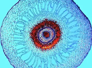
Background imageSectioned Collection: Water fern rhizome, light micrograph

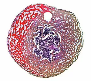
Water fern rhizome, light micrograph
Water fern rhizome. Polarised light micrograph of a section through a rhizome from a water fern (Marselia vestita). Under the epidermis (the outer layer)

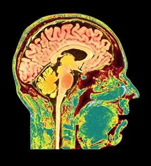
Background imageSectioned Collection: Normal human brain, MRI scan C016 / 8850

Normal human brain, MRI scan C016 / 8850
Brain. Coloured magnetic resonance imaging (MRI) scan of a sagittal section through a patients head showing a healthy human brain

Background imageSectioned Collection: Ovarian follicle, light micrograph C016 / 0519

Ovarian follicle, light micrograph C016 / 0519
Ovarian follicle. Light micrograph of a section through a secondary ovarian follicle showing signs of maturation into the antral follicle class

Background imageSectioned Collection: Purkinje nerve cell, TEM C014 / 0583

Purkinje nerve cell, TEM C014 / 0583
Purkinje nerve cell. Transmission electron micrograph (TEM) of a purkinje nerve cell (green) from the cerebellum of the brain, showing the cell body (centre) and its primary dendrite (cell process)

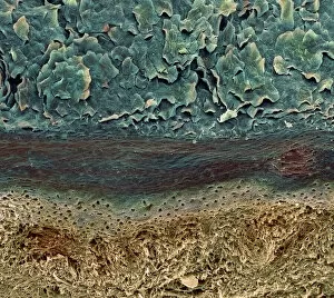
Background imageSectioned Collection: Skin layers, SEM

Skin layers, SEM

Background imageSectioned Collection: Pancreatic islet of Langerhans

Pancreatic islet of Langerhans
Islet of Langerhans. Light micrograph of a section through an islet of Langerhans (pale, centre) in pancreas tissue. This clump of secretory cells forms part of the endocrine system of the body

Background imageSectioned Collection: Blood vessel

Blood vessel. Coloured scanning electron micrograph (SEM) of a section through a blood vessel in the skin. The vessel contains red blood cells (erythrocytes, red). Magnification unknown

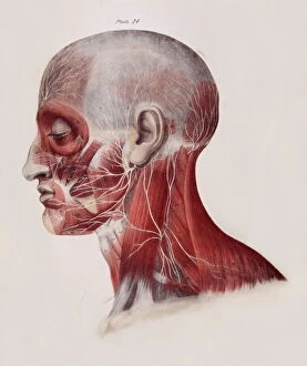
Background imageSectioned Collection: Facial nerves

Facial nerves. Historical anatomical artwork of a side view of a dissected human head showing the nerves (white) and muscles (red) of the face and neck
Background imageSectioned Collection: Osteoporotic bone

Osteoporotic bone

Background imageSectioned Collection: Intestinal microvilli, TEM

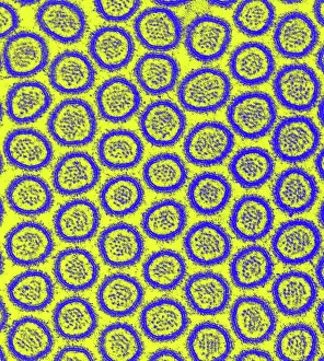
Intestinal microvilli, TEM
Intestinal microvilli, coloured transmission electron micrograph (TEM). The microvilli are finger-like projections (seen here in circular cross-section)

Background imageSectioned Collection: Human tooth anatomy, artwork

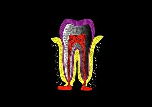
Human tooth anatomy, artwork
Human tooth anatomy. Artwork of a cross-section through a human tooth to show its anatomical structure. The two main areas are the crown (above the gum-line) and the root (embedded in the gum)

Background imageSectioned Collection: Maiden hair tree stem, SEM

Maiden hair tree stem, SEM
Maiden hair tree stem. Coloured scanning electron micrograph (SEM) of a section through a maidenhair tree (Ginkgo biloba) stem. At centre is the pith, which consists of soft, spongy parenchyma cells

Background imageSectioned Collection: Tomato leaf, light micrograph

Tomato leaf, light micrograph
Tomato leaf. Light micrograph of a transverse section through the midrib of a tomato (Lycopersicum esculentum) leaf. The upper and lower epidermis on the surfaces of the leaf are blue

Background imageSectioned Collection: Xylem tissue, SEM

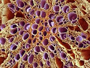
Xylem tissue, SEM
Xylem tissue. Coloured scanning electron micrograph (SEM) of a section through xylem tissue from a dicotyledon rootlet. Xylem vessels (purple)

Background imageSectioned Collection: Wood, light micrograph

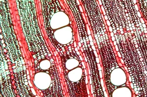
Wood, light micrograph
Wood. Light micrograph of a transverse section through wood showing annual growth rings (near horizontal bands). A years growth consists of a porous band, which is the slow winter growth

Background imageSectioned Collection: Spongy bone, light micrograph

Spongy bone, light micrograph
Spongy bone. Light micrograph of a section through stained and decalcified human spongy bone. This bone type is also called cancellous bone

Background imageSectioned Collection: Heart muscle, confocal light micrograph

Heart muscle, confocal light micrograph
Heart muscle. Confocal light micrograph of a section through cardiac (heart) muscle. Cardiac muscle consists of branching elongated muscle cells

Background imageSectioned Collection: Artistic view of bare tree in sections, reflecting over water

Artistic view of bare tree in sections, reflecting over water

Background imageSectioned Collection: Close-up of the texture of a tree

Close-up of the texture of a tree

Background imageSectioned Collection: Different color variations of a tree trunk with fern

Different color variations of a tree trunk with fern

Background imageSectioned Collection: Animal cell structure

Animal cell structure. Artwork of a section through an animal cell. At lower centre is the nucleus, which contains the cells genetic information in the form of DNA (deoxyribonucleic acid)

Background imageSectioned Collection: The Rake in Bedlam, plate VIII from A Rakes Progress, 1763 (engraving)

The Rake in Bedlam, plate VIII from A Rakes Progress, 1763 (engraving)
GHL206154 The Rake in Bedlam, plate VIII from A Rakes Progress, 1763 (engraving) by Hogarth, William (1697-1764); length: 56 cm; London Metropolitan Archives

Background imageSectioned Collection: SOCEMA TGA-01 turboprop at the Paris Salon Aeronautique 1949

SOCEMA TGA-01 turboprop at the Paris Salon Aeronautique 1949
SOCEMA TGA-01 turboprop at the XVIIIeme Salon Aeronautique du Paris 1949 at the Grand Palais in central Paris, (flying demonstrations were given at Orly Airport)

Background imageSectioned Collection: Normal human brain, MRI scan C016 / 8843

Normal human brain, MRI scan C016 / 8843
Brain. Coloured magnetic resonance imaging (MRI) scan of a sagittal section through a healthy human brain. The highly folded area (brown) is the cerebrum, which is responsible for conscious thought

Background imageSectioned Collection: Human olfactory system, artwork C016 / 9378

Human olfactory system, artwork C016 / 9378
Human olfactory system. Computer artwork of a section through a human head, showing the olfactory receptor cells (brush-like), and their location in the olfactory bulb (sausage-shape)

Background imageSectioned Collection: Chloroplast, SEM

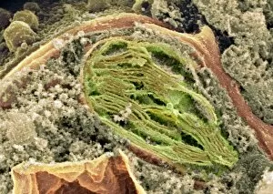
Chloroplast, SEM
Chloroplast. Coloured scanning electron micrograph (SEM) of a section through a plant cell, showing a fractured chloroplast (green)

Background imageSectioned Collection: Sectioned skull in profile to left with the left side of the cranium removed, 1651

Sectioned skull in profile to left with the left side of the cranium removed, 1651

Background imageSectioned Collection: Close-up of puppy lying on sand

Close-up of puppy lying on sand

Background imageSectioned Collection: A sectioned Pratt & Whitney R-4360 Wasp Major radial

A sectioned Pratt & Whitney R-4360 Wasp Major radial

Background imageSectioned Collection: Intestinal villi, SEM

Intestinal villi, SEM
Intestinal villi. Coloured scanning electron micrograph (SEM) of a section through a part of the small intestine, showing the villi that cover its inner surface

Background imageSectioned Collection: Cervix, light micrograph F006 / 9805

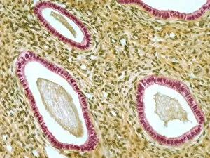
Cervix, light micrograph F006 / 9805
Cervix. Light micrograph of a section though the mucosa of the cervix, the neck of the uterus. The purple-lined areas are cervical glands, which produce mucus (pink inside the glands)

Background imageSectioned Collection: Fallopian tube, light micrograph F006 / 9799

Fallopian tube, light micrograph F006 / 9799
Fallopian tube. Light micrograph of a cross-section through a fallopian tube. The lumen (white) is seen, along with mucosal folds lined with simple columnar ciliated epithelium (purple)

Background imageSectioned Collection: Thymus gland, light micrograph C015 / 4970

Thymus gland, light micrograph C015 / 4970
Thymus gland. Light micrograph of a section through tissue from the thymus gland, part of the lymphoid system. The dense areas are the thymic cortex, which produces lots of lymphocytes

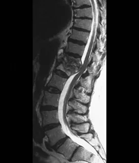
Background imageSectioned Collection: Tuberculosis of the spine, MRI scan

Tuberculosis of the spine, MRI scan
Tuberculosis of the spine. Magnetic resonance imaging (MRI) scan of a sagittal section through the spine of a 74-year-old patient with tuberculosis (TB) of the spine, or Potts disease

Background imageSectioned Collection: Spondylolisthesis, 3D CT scan

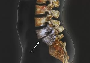
Spondylolisthesis, 3D CT scan
Spondylolisthesis. Coloured 3D computed tomography (CT) scan of a section through the lumbar (lower back) spine of a 39-year-old patient with spondylolisthesis

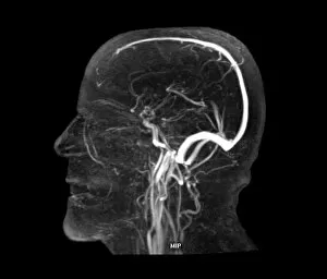
Background imageSectioned Collection: Vascular system of the head, MRI scan

Vascular system of the head, MRI scan
Vascular system of the head. Magnetic resonance imaging (MRI) angiogram of a sagittal section through the head of a healthy 43-year-old, showing the blood vessels (white) of the brain and neck

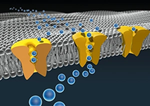
Background imageSectioned Collection: Cell membrane ion channels, artwork C016 / 7689

Cell membrane ion channels, artwork C016 / 7689
Cell membrane ion channels. Computer artwork of a section through the membrane of an animal cell, showing transmembrane ion channel proteins (yellow)

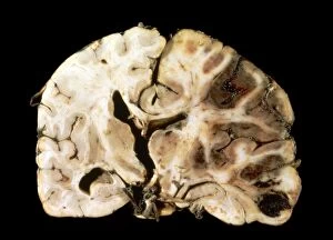
Background imageSectioned Collection: Brain injury C016 / 8920

Brain injury C016 / 8920
Brain injury. Gross specimen of a section through a brain showing the effects of a blunt trauma. The hemisphere at right is showing extensive haemorrhaging (bleeding)

Background imageSectioned Collection: Eosinophil white blood cell, TEM

Eosinophil white blood cell, TEM
Eosinophil white blood cell. Transmission electron micrograph (TEM) of a section through an eosinophil white blood cell (leukocyte)

Background imageSectioned Collection: Pancreatic acinar cell

Pancreatic acinar cell. Transmission electron micrograph (TEM) of a section through an enzyme-secreting acinar cell in the human pancreas, showing part of the nucleus (round, far left)

Background imageSectioned Collection: Hepatocyte liver cell, TEM

Hepatocyte liver cell, TEM
Hepatocyte liver cell. Transmission electron micrograph (TEM) of a section through an hepatocyte liver cell, showing rough and smooth endoplasmic reticulum (ER, dark lines)

Background imageSectioned Collection: Liver tissue, TEM

Liver tissue, TEM
Liver tissue. Transmission electron micrograph (TEM) of a section through the liver, showing part of a radial cord of hepatocyte liver cells (dark) and the vascular sinusoids (white)

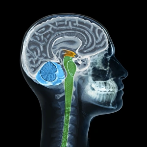
Background imageSectioned Collection: Human head, X-ray artwork

Human head, X-ray artwork
Human head. Computer artwork of an X-ray through a human head, showing the brain (grey, top), cerebellum (blue), and brainstem and spinal cord (green)

Background imageSectioned Collection: Trachea lining, SEM C015 / 9936

Trachea lining, SEM C015 / 9936
Trachea lining. Coloured scanning electron micrograph (SEM) of a section through the lining of the trachea (wind pipe). The trachea links the larynx (voice box) to the lungs

Background imageSectioned Collection: Vagus nerve anatomy, artwork C018 / 0300

Vagus nerve anatomy, artwork C018 / 0300
Vagus nerve anatomy. Artwork of a human body from the side, showing the internal anatomy and the location of the vagus nerve (yellow)

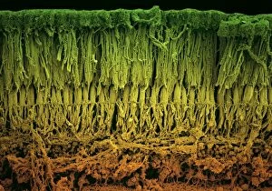
Background imageSectioned Collection: Retina layers, SEM C018 / 0520

Retina layers, SEM C018 / 0520
Retina layers, coloured scanning electron micrograph (SEM). The retina of the human eye is found on the inside of the eyeball and is formed from a number of layers

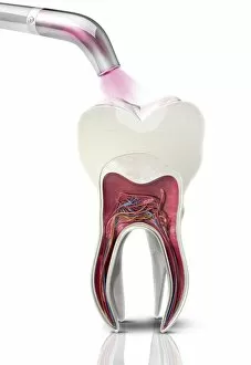
Background imageSectioned Collection: Dental filling polymerisation, artwork C014 / 2025

Dental filling polymerisation, artwork C014 / 2025
Dental filling polymerisation. Artwork of a cross-section of a human tooth, with a filling (white) being set using an ultraviolet light lamp tool


All Professionally Made to Order for Quick Shipping

"Exploring the Intricacies: From Cerebellum Tissue to Fusion Research, Unveiling the World Sectioned" Delving into the depths of scientific exploration, a light micrograph reveals the intricate structure of cerebellum tissue. Witnessing a Higgs boson event through the lens of ATLAS detector C013/6892, scientists capture an awe-inspiring moment in particle physics. A meticulously crafted model showcases the complexity and beauty of the human brain, inviting us to marvel at its wonders. Peering into hippocampus brain tissue under a microscope, we uncover secrets that may hold keys to memory formation and learning processes. In pursuit of limitless energy sources, fusion research takes center stage as we examine a tokamak device designed for controlled nuclear fusion reactions. The inner workings of our brains come alive through an MRI scan, revealing detailed brain anatomy like never before imagined. Illuminated by light microscopy, dicotyledon plant stem sections unveil nature's architectural masterpiece in stunning detail. EDTA crystals take on mesmerizing patterns when viewed under intense magnification - showcasing their unique molecular arrangement through light micrography. Journeying deep within our bodies, kidney tubules are sectioned to shed light on their vital role in maintaining fluid balance and waste removal systems. An X-ray image exposes every contour and intricacy of a human skull – offering glimpses into our skeletal framework with remarkable clarity. Through transmission electron microscopy (TEM), rough endoplasmic reticulum emerges as an interconnected network responsible for protein synthesis within cells' cytoplasmic matrix Computer artwork visualizes Alzheimer's-affected brain regions – reminding us why continued research is crucial in unraveling this devastating disease. In this captivating journey across various scientific disciplines and microscopic landscapes, "sectioned" unveils hidden worlds that expand our understanding of the universe and ourselves.