Fraud Blocker Skip to main content

Secondary Structure Collection

Secondary structure refers to the intricate folding patterns that proteins and nucleic acids adopt, playing a crucial role in their functionality

Background imageSecondary Structure Collection: DNA transcription, molecular model

DNA transcription, molecular model. Secondary structure of the enzyme RNA polymerase II synthesising a mRNA (messenger ribonucleic acid, lilac) strand from a DNA (deoxyribonucleic acid)

Background imageSecondary Structure Collection: Anaesthetic inhibiting an ion channel C015 / 6718

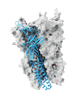
Anaesthetic inhibiting an ion channel C015 / 6718
Anaesthetic inhibiting an ion channel. Computer model showing the structure of propofol anaesthetic drug molecules (spheres)

Background imageSecondary Structure Collection: Secondary structure of proteins, artwork

Secondary structure of proteins, artwork
Secondary structure of proteins, computer artwork. The secondary structure is the shape taken by the strands of proteins, which are biological polymers of amino acids

Background imageSecondary Structure Collection: Nucleosome molecule

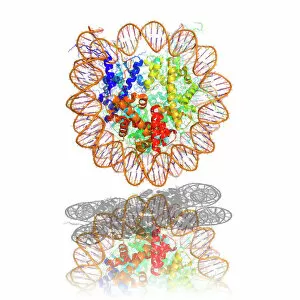
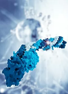
Nucleosome molecule, computer model. A nucleosome is a subunit of chromatin, the substance that forms chromosomes. It consists of a short length of DNA (deoxyribonucleic acid)

Background imageSecondary Structure Collection: DNA nucleosome, molecular model

DNA nucleosome, molecular model
DNA nucleosome. Molecular model of a nucleosome, the fundamental repeating unit used to package DNA (deoxyribonucleic acid) inside cell nuclei

Background imageSecondary Structure Collection: Bacterial ribosome

Bacterial ribosome. Computer model showing the secondary structure of a 30S (small) ribosomal sub-unit from the bacteria Thermus thermophilus

Background imageSecondary Structure Collection: HIV reverse transcription enzyme

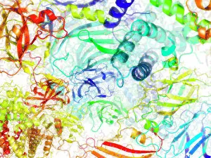
HIV reverse transcription enzyme. Molecular models of the reverse transcriptase enzyme found in HIV (the human immunodeficiency virus)

Background imageSecondary Structure Collection: Hepatitis C virus enzyme, molecular model

Hepatitis C virus enzyme, molecular model
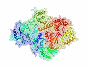
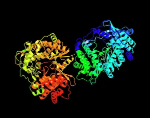
Hepatitis C virus enzyme. Molecular model of a genetic enzyme from the Hepatitis C virus. This enzyme is called HC-J4 RNA polymerase

Background imageSecondary Structure Collection: Interferon molecule

Interferon molecule. Computer model showing the secondary structure of a molecule of interferon. Interferons are proteins produced by white blood cells as part of the immune response to invading

Background imageSecondary Structure Collection: Coagulation factor complex molecule C014 / 0139

Coagulation factor complex molecule C014 / 0139
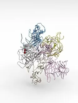
Coagulation factor complex molecule. Molecular model showing the interaction between coagulation factor VIII (FVIII, pink, blue and yellow) and factor IXa (FIXa, cream and grey)

Background imageSecondary Structure Collection: Ghrelin hormone molecule

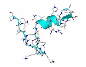
Ghrelin hormone molecule. Computer model showing the crystal structure of the human hormone ghrelin. The crystal structure consists of both the secondary structure

Background imageSecondary Structure Collection: Tumour suppressor protein and DNA C017 / 3647

Tumour suppressor protein and DNA C017 / 3647
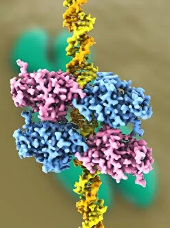
Tumour suppressor protein and DNA. Computer artwork showing a molecule of the tumour suppressor protein p53 (blue and pink) bound to a molecule of DNA (deoxyribonucleic acid, yellow and orange)

Background imageSecondary Structure Collection: Antibiotic resistance enzyme molecule C017 / 2272

Antibiotic resistance enzyme molecule C017 / 2272
Antibiotic resistance enzyme. Molecular model of the New Delhi metallo-beta-lactamase 1 enzyme. This bacterial enzyme confers antibiotic resistance on cells that carry it

Background imageSecondary Structure Collection: Antibiotic resistance enzyme molecule C017 / 2271

Antibiotic resistance enzyme molecule C017 / 2271
Antibiotic resistance enzyme. Molecular model of the New Delhi metallo-beta-lactamase 1 enzyme. This bacterial enzyme confers antibiotic resistance on cells that carry it

Background imageSecondary Structure Collection: Sirtuin enzyme and p53, artwork C017 / 3659

Sirtuin enzyme and p53, artwork C017 / 3659
Sirtuin enzyme and p53. Computer artwork of a sirtuin (Sir2) enzyme (pink) bound to a p53 peptide (orange). Sir2 enzymes form a unique class of NAD(+)

Background imageSecondary Structure Collection: Tumour suppressor protein and DNA C017 / 3644

Tumour suppressor protein and DNA C017 / 3644
Tumour suppressor protein and DNA. Computer artwork showing a molecule of the tumour suppressor protein p53 (blue and pink) bound to a molecule of DNA (deoxyribonucleic acid, yellow and orange)

Background imageSecondary Structure Collection: Ricin molecule, artwork C017 / 3651

Ricin molecule, artwork C017 / 3651
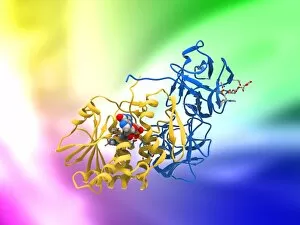
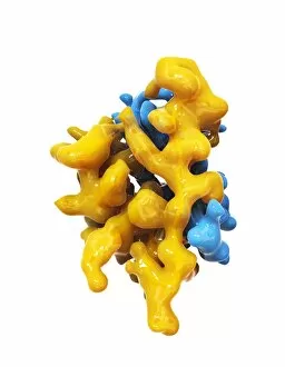
Ricin molecule. Computer artwork showing the structure of a molecule of the toxic protein ricin. Ricin comprises two entwined amino acid chains; A (yellow) and B (blue)

Background imageSecondary Structure Collection: Ricin molecule, artwork C017 / 3650

Ricin molecule, artwork C017 / 3650
Ricin molecule. Computer artwork showing the structure of a molecule of the toxic protein ricin. Ricin comprises two entwined amino acid chains; A (yellow) and B (blue)

Background imageSecondary Structure Collection: Sirtuin enzyme and p53, artwork C017 / 3658

Sirtuin enzyme and p53, artwork C017 / 3658
Sirtuin enzyme and p53. Computer artwork of a sirtuin (Sir2) enzyme (pink) bound to a p53 peptide (orange). Sir2 enzymes form a unique class of NAD(+)

Background imageSecondary Structure Collection: SIRT3 molecule, artwork C017 / 3657

SIRT3 molecule, artwork C017 / 3657
SIRT3 molecule. Computer artwork showing the structure of a molecule of NAD-dependent deacetylase sirtuin-3, mitochondrial (SIRT3)

Background imageSecondary Structure Collection: Tumour suppressor protein and DNA C017 / 3646

Tumour suppressor protein and DNA C017 / 3646
Tumour suppressor protein and DNA. Computer artwork showing a molecule of the tumour suppressor protein p53 (blue and pink) bound to a molecule of DNA (deoxyribonucleic acid, yellow and orange)

Background imageSecondary Structure Collection: Fibrinogen molecule C014 / 0473

Fibrinogen molecule C014 / 0473
Fibrinogen. Molecular model showing the structure of the blood clotting glycoprotein fibrinogen (factor I). The molecule consists of two sets of alpha (grey)

Background imageSecondary Structure Collection: Tumour suppressor protein and DNA C017 / 3645

Tumour suppressor protein and DNA C017 / 3645
Tumour suppressor protein and DNA. Computer artwork showing a molecule of the tumour suppressor protein p53 (blue and pink) bound to a molecule of DNA (deoxyribonucleic acid, yellow and orange)

Background imageSecondary Structure Collection: EcoRV restriction enzyme molecule C014 / 2117

EcoRV restriction enzyme molecule C014 / 2117
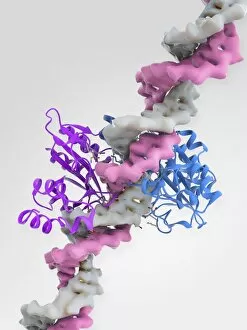
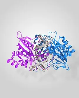
EcoRV restriction enzyme. Molecular model of the type II restriction enzyme EcoRV (purple and blue) bound to a DNA molecule (deoxyribonucleic acid, pink and white)

Background imageSecondary Structure Collection: Sirtuin enzyme and p53, artwork C017 / 3660

Sirtuin enzyme and p53, artwork C017 / 3660
Sirtuin enzyme and p53. Computer artwork of a sirtuin (Sir2) enzyme (blue) bound to a p53 peptide (pink). Sir2 enzymes form a unique class of NAD(+)

Background imageSecondary Structure Collection: EcoRV restriction enzyme molecule C014 / 2114

EcoRV restriction enzyme molecule C014 / 2114
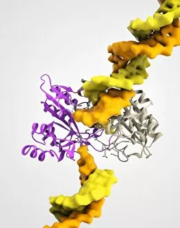
EcoRV restriction enzyme. Molecular model of the type II restriction enzyme EcoRV (white and gold) bound to a cleaved section of DNA (deoxyribonucleic acid, orange and yellow)

Background imageSecondary Structure Collection: Ricin molecule, artwork C017 / 3649

Ricin molecule, artwork C017 / 3649
Ricin molecule. Computer artwork showing the structure of a molecule of the toxic protein ricin. Ricin comprises two entwined amino acid chains; A (yellow) and B (blue)

Background imageSecondary Structure Collection: Insulin molecule C014 / 2121

Insulin molecule C014 / 2121
Insulin molecule. Molecular module of insulin showing its secondary structure. Insulin is a hormone produced by the pancreas

Background imageSecondary Structure Collection: EcoRV restriction enzyme molecule C014 / 2115

EcoRV restriction enzyme molecule C014 / 2115
EcoRV restriction enzyme. Molecular model of the type II restriction enzyme EcoRV (purple and blue) bound to a DNA molecule (deoxyribonucleic acid, pink and white)

Background imageSecondary Structure Collection: Blood clotting factor VIII molecule C014 / 0406

Blood clotting factor VIII molecule C014 / 0406
Blood clotting factor VIII, molecular model showing the secondary structure. Factor VIII, also known as anti-haemophilic factor, is an essential blood clotting protein in humans

Background imageSecondary Structure Collection: Nucleosome molecule F006 / 9323

Nucleosome molecule F006 / 9323
Nucleosome, molecular model. A nucleosome is a subunit of chromatin, the substance that forms chromosomes. It consists of a short length of DNA (deoxyribonucleic acid)

Background imageSecondary Structure Collection: Nucleosome molecule F006 / 9314

Nucleosome molecule F006 / 9314
Nucleosome, molecular model. A nucleosome is a subunit of chromatin, the substance that forms chromosomes. It consists of a short length of DNA (deoxyribonucleic acid)

Background imageSecondary Structure Collection: Leptin molecule F006 / 9243

Leptin molecule F006 / 9243
Leptin molecule. Molecular model of the human hormone leptin. Leptin is a protein produced by adipose (fat) tissue. It interacts with receptors in the brains hypothalamus to signal when a person is

Background imageSecondary Structure Collection: Nucleosome molecule F006 / 9235

Nucleosome molecule F006 / 9235
Nucleosome, molecular model. A nucleosome is a subunit of chromatin, the substance that forms chromosomes. It consists of a short length of DNA (deoxyribonucleic acid)

Background imageSecondary Structure Collection: Interleukin-6, molecular model F006 / 9232

Interleukin-6, molecular model F006 / 9232
Interleukin-6. Molecular model of the cytokine protein human interleukin-6. This protein is produced in the body and has a wide variety of functions in the immune system

Background imageSecondary Structure Collection: MUC5B molecule

MUC5B molecule. Computer model showing the structure of a molecule of the protein MUC5B (mucin 5 subtype B). Mucins key characteristic is their ability to form gels

Background imageSecondary Structure Collection: Myoglobin protein, molecular model C016 / 6575

Myoglobin protein, molecular model C016 / 6575
Myoglobin protein. Molecular model showing the structure of the myoglobin protein. Myoglobin is a protein found in muscle tissue

Background imageSecondary Structure Collection: Bicalutamide drug binding to receptor

Bicalutamide drug binding to receptor. Molecular model of the drug bicalutamide (lower right) binding to a receptor protein (ribbons)

Background imageSecondary Structure Collection: Nobel Prize, conceptual artwork

Nobel Prize, conceptual artwork
Nobel Prize. Conceptual artwork representing the Nobel Prizes, leading awards made annually for outstanding cultural and scientific advances

Background imageSecondary Structure Collection: Bacterial ribosome and protein synthesis

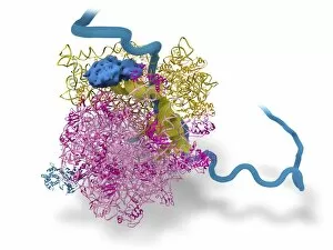
Bacterial ribosome and protein synthesis. Molecular model showing a bacterial ribosome reading an mRNA (messenger ribonucleic acid) strand (blue) and synthesising a protein

Background imageSecondary Structure Collection: Ricin molecule, artwork C017 / 3656

Ricin molecule, artwork C017 / 3656
Ricin molecule. Computer artwork showing the structure of a molecule of the toxic protein ricin. Ricin comprises two entwined amino acid chains; A (yellow) and B (blue)

Background imageSecondary Structure Collection: Ricin molecule, artwork C017 / 3655

Ricin molecule, artwork C017 / 3655
Ricin molecule. Computer artwork showing the structure of a molecule of the toxic protein ricin. Ricin comprises two entwined amino acid chains; A (yellow) and B (blue)

Background imageSecondary Structure Collection: Blood clotting factor VIII molecule C014 / 0407

Blood clotting factor VIII molecule C014 / 0407
Blood clotting factor VIII, molecular model showing the secondary structure. Factor VIII, also known as anti-haemophilic factor, is an essential blood clotting protein in humans

Background imageSecondary Structure Collection: Coagulation factor complex molecule C014 / 0409

Coagulation factor complex molecule C014 / 0409
Coagulation factor complex molecule. Molecular model showing the interaction between coagulation factor VIII (FVIII, pink, blue and yellow), factor IXa (FIXa)

Background imageSecondary Structure Collection: Insulin molecule C014 / 2122

Insulin molecule C014 / 2122
Insulin, molecular module. Insulin is a hormone produced by the pancreas. It consists of two peptide chains, A (blue) and B (yellow), which are linked by disulphide bridges

Background imageSecondary Structure Collection: Blood clotting factor VIII molecule C014 / 0408

Blood clotting factor VIII molecule C014 / 0408
Blood clotting factor VIII, molecular model. Factor VIII, also known as anti-haemophilic factor, is an essential blood clotting protein in humans

Background imageSecondary Structure Collection: EcoRV restriction enzyme molecule C014 / 2113

EcoRV restriction enzyme molecule C014 / 2113
EcoRV restriction enzyme. Molecular model of the type II restriction enzyme EcoRV (pink and blue) bound to a cleaved section of DNA (deoxyribonucleic acid, white)

Background imageSecondary Structure Collection: EcoRV restriction enzyme molecule C014 / 2118

EcoRV restriction enzyme molecule C014 / 2118
EcoRV restriction enzyme. Molecular model of the type II restriction enzyme EcoRV (purple and beige) bound to a DNA molecule (deoxyribonucleic acid, yellow and orange)


All Professionally Made to Order for Quick Shipping

Secondary structure refers to the intricate folding patterns that proteins and nucleic acids adopt, playing a crucial role in their functionality. In one captivating image, an anaesthetic inhibits an ion channel (C015/6718), highlighting how secondary structure impacts cellular processes. Another snapshot showcases DNA transcription, unveiling the molecular model of this essential process. Artwork depicting the secondary structure of proteins captivates our imagination as we marvel at the intricacy and beauty within each fold. The nucleosome molecule further emphasizes this complexity, showcasing how DNA wraps around histone proteins to form a compact structure. The bacterial ribosome stands tall as a testament to secondary structures' significance in protein synthesis. Meanwhile, the HIV reverse transcription enzyme reminds us of its vital role in converting viral RNA into DNA during infection. Molecular models provide insight into hepatitis C virus enzymes and interferon molecules—both critical players in disease progression and immune response modulation. Similarly, human growth hormone molecules hold immense importance for development and metabolism regulation. Exploring coagulation factor complex molecules (C014/0139) unravels mechanisms behind blood clotting—a process dependent on precise secondary structures working together seamlessly. Ghrelin hormone molecules intrigue us with their involvement in appetite regulation and energy balance maintenance. From anaesthetics influencing ion channels to hormones orchestrating bodily functions, understanding secondary structures unlocks countless mysteries within biological systems. These captivating images remind us of the intricate dance occurring at a microscopic level—the delicate folds dictating life's grand symphony.