
Scanning Collection (page 8)
"Unveiling the Secrets: Scanning the Unseen World" Step into a world of discovery as we embark on a journey of scanning


All Professionally Made to Order for Quick Shipping
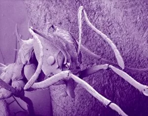
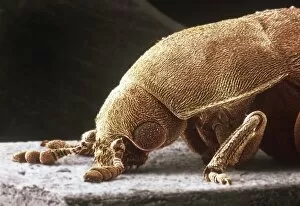
"Unveiling the Secrets: Scanning the Unseen World" Step into a world of discovery as we embark on a journey of scanning, revealing hidden wonders and unraveling mysteries. From the towering Old Hartlepool Lighthouse in north east England to cutting-edge fingerprint scanners, our quest for knowledge knows no bounds. Intriguingly, even nature's creations hold surprises. Picture a young German Shepherd Dog lying on lush green grass, its innocent gaze inviting us to explore further. Delving deeper, we encounter snail teeth - microscopic marvels that defy expectations with their intricate structure. But our exploration doesn't stop there; we venture into realms unseen by the naked eye. Enter Plasmodium sp. , the malarial parasite that has plagued humanity for centuries. Through advanced scanning techniques, scientists strive to understand this elusive foe and develop effective treatments. As we continue our voyage through images like Picture No. 11675478 and Picture No. 11675612, breathtaking landscapes unfold before us. Each scan captures moments frozen in time - from serene sunsets painting the sky with vibrant hues to majestic mountains standing tall against an azure backdrop. Yet not all scans are purely aesthetic; some serve vital purposes beyond aesthetics alone. Take Aspergillus - an organism whose presence can pose health risks if left undetected. By employing sophisticated scanning methods, experts can identify potential threats and safeguard public health. Our historical expedition takes us back in time to witness technological advancements firsthand - X-ray security machines from 1900 paving the way for modern-day safety measures at airports worldwide (Picture No. 10876997). These early pioneers laid foundations upon which today's state-of-the-art scanners stand tall. With each scan captured in Picture No. 11675582 or any other frame along this captivating journey, we realize that "scanning" is more than just a process; it is an art form that allows us to peer into the intricate details of our world, both seen and unseen.