Fraud Blocker Skip to main content

Scanning Electron Micrograph Collection (page 2)

"Exploring the Microscopic World

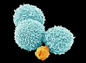
Background imageScanning Electron Micrograph Collection: White blood cells, SEM

White blood cells, SEM
White blood cells. Coloured scanning electron micrograph (SEM) of white blood cells (leucocytes). Magnification: x2, 400 when printed at 10 centimetres wide

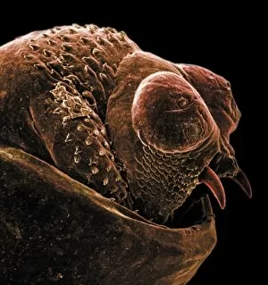
Background imageScanning Electron Micrograph Collection: The anus of a bot fly

The anus of a bot fly
Scanning electron microscope image of the anus of a bot fly. Image on display in the Darwin Centre at the Natural History Museum, London

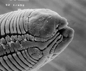
Background imageScanning Electron Micrograph Collection: Roundworm

Roundworm
Scanning electron microscope (SEM) image of a parasitic roundworms head (x 1000 on a standard 9 cm wide print)

Background imageScanning Electron Micrograph Collection: Moth egg

Moth egg
Scanning electron microscope (SEM) image of a moth egg (x 90). The caterpillar emerges by chewing through the shell

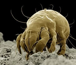
Background imageScanning Electron Micrograph Collection: Dermatophagoides sp. dust mite

Dermatophagoides sp. dust mite
Scanning electron microscope image of a dust mite. Dust mites are secondary to pollen as a cause for allergies and they live in bedding, soft furniture and carpets

Background imageScanning Electron Micrograph Collection: Sarcoptes scabiei, scabies mite

Sarcoptes scabiei, scabies mite
Scanning electron microscope image of an itch or scabies mite, a parasite that infests a wide variety of mammalian hosts including man

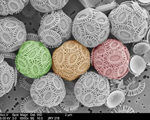
Background imageScanning Electron Micrograph Collection: Emiliania huxleyi coccolithophores

Emiliania huxleyi coccolithophores collected from a bloom in the SW Approaches to the English Channel in June 2004. Date: 2004

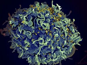
Background imageScanning Electron Micrograph Collection: Scanning electron micrograph of HIV particles infecting a human H9 T cell

Scanning electron micrograph of HIV particles infecting a human H9 T cell

Background imageScanning Electron Micrograph Collection: Coxiella burnetii, the bacteria that causes Q Fever

Coxiella burnetii, the bacteria that causes Q Fever. A dry fracture of a Vero cell exposing the contents of a vacuole where Coxiella burnetii are busy growing

Background imageScanning Electron Micrograph Collection: Sea salt

Sea salt
A scanning electron microscope (SEM) image of sea salt, artificially coloured by computer

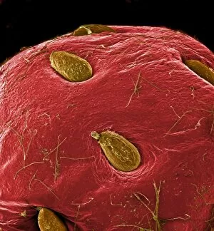
Background imageScanning Electron Micrograph Collection: Fragaria sp. strawberry

Fragaria sp. strawberry
A scanning electron microscope (SEM) image of a strawberry (Fragaria sp.), artificially coloured by computer

Background imageScanning Electron Micrograph Collection: Tyrophagus casei, cheese mite

Tyrophagus casei, cheese mite
Scanning electron microscope image of a cheese mite (x 170). These creatures are generally considered to be a pest, however they are added to Altenburger cheese to give it flavour

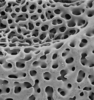
Background imageScanning Electron Micrograph Collection: SEM of echinoderm steroem

SEM of echinoderm steroem
A SEM of an example of stereom of an echinoderm (phylum which consists of 5 classes including starfish). Stereom is the structure formed by the fine networks of calcium carbonate which constitute

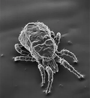
Background imageScanning Electron Micrograph Collection: Dermanyssus gallinae, red or poultry mite

Dermanyssus gallinae, red or poultry mite
Scanning electron microscope image of the red or poutry mite. Adults appear red when engorged with blood, but otherwise are black, grey or white. Females are about 1mm long

Background imageScanning Electron Micrograph Collection: Phthiracarus sp. box mite or armadillo mite

Phthiracarus sp. box mite or armadillo mite
Scanning electron microscope (SEM) image of a box mite, showing how the body has fused into one single segment

Background imageScanning Electron Micrograph Collection: Ceratodon purpureus, ceratodon moss spore capsule

Ceratodon purpureus, ceratodon moss spore capsule
Scanning electron microscope (SEM) image of a ceratodon moss spore capsule (x 650 on a standard 9 cm wide print)

Background imageScanning Electron Micrograph Collection: Foraminifer

Foraminifer
Scanning electron microscope (SEM) image of a foraminifer - a single celled organism

Background imageScanning Electron Micrograph Collection: Dermatophagoides pteronyssius, dust mite

Dermatophagoides pteronyssius, dust mite
Scanning electron microscope image showing a dust mite (x 250 on standard 9cm wide print). This image has been artificially coloured by a computer

Background imageScanning Electron Micrograph Collection: Marine Diatoms

Marine Diatoms
LRMC-3 Scanning Electron Micrograph (SEM): Marine Diatoms Magnification x2400 (when printed A4, 29.7 cm cm wide) David McCarthy and Dae Sasitorn / ardea.com Last Refuge contact details

Background imageScanning Electron Micrograph Collection: Silicoflagellate

Silicoflagellate
LRDS-297 Scanning Electron Micrograph (SEM): Silicoflagellate, Skeleton bearing stage of Dictyocha speculum Magnification x3

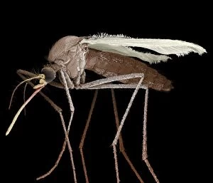
Background imageScanning Electron Micrograph Collection: Scanning Electron Micrograph (SEM): Mosquito, Magnification x 35 (A4 size: 29. 7 cm width)

Scanning Electron Micrograph (SEM): Mosquito, Magnification x 35 (A4 size: 29. 7 cm width)
LRDS-84 Mosquito Scanning Electron Micrograph (SEM) Culex pipiens Magnification x 35 (A4 size: 29.7 cm width) Coloured by hand to enhance natural features

Background imageScanning Electron Micrograph Collection: Scanning Electron Micrograph (SEM): Flour Mite, Magnification x 500 (A4 size: 29. 7 cm width)

Scanning Electron Micrograph (SEM): Flour Mite, Magnification x 500 (A4 size: 29. 7 cm width)
LRDS-58 Flour Mite Scanning Electron Micrograph (SEM) Acarus siro Magnification x 500 (A4 size: 29.7 cm width) Coloured by hand to enhance natural features

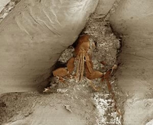
Background imageScanning Electron Micrograph Collection: Scanning Electron Micrograph (SEM): Cat Flea, Magnification x250 (A4 size: 29. 7 cm width)

Scanning Electron Micrograph (SEM): Cat Flea, Magnification x250 (A4 size: 29. 7 cm width)
LRDS-54 Cat Flea Scanning Electron Micrograph (SEM) Ctenocephalides felis Magnification x250 (A4 size: 29.7 cm width) Coloured by hand to enhance natural features

Background imageScanning Electron Micrograph Collection: Scanning Electron Micrograph (SEM): Chigoe Flea, Magnification x 200 (A4 size: 29. 7 cm width)

Scanning Electron Micrograph (SEM): Chigoe Flea, Magnification x 200 (A4 size: 29. 7 cm width)
LRDS-137 Chigoe Flea Scanning Electron Micrograph (SEM) Tunga penetrans Magnification x 200 (A4 size: 29.7 cm width) Coloured by hand to enhance natural features

Background imageScanning Electron Micrograph Collection: Scanning Electron Micrograph (SEM): Springtail, Order Collembola; Magnification x 150 (A4 size)

Scanning Electron Micrograph (SEM): Springtail, Order Collembola; Magnification x 150 (A4 size: 29. 7 cm width)
LRDS-122 Springtail, Order Collembola Scanning Electron Micrograph (SEM) Magnification x 150 (A4 size: 29.7 cm width) Coloured by hand to enhance natural features

Background imageScanning Electron Micrograph Collection: Chrysanthemum, CT scan image

Chrysanthemum, CT scan image
CT Scan image of a Chrysanthemum

Background imageScanning Electron Micrograph Collection: Syracosphaera anthos

Syracosphaera anthos
Coccosphere from the Western Mediterranean. False coloured to show the shell is formed of inner and outer layers of coccoliths with very different structure

Background imageScanning Electron Micrograph Collection: Colorized image of HIV-infected H9 T-cell

Colorized image of HIV-infected H9 T-cell

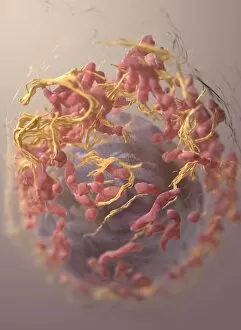
Background imageScanning Electron Micrograph Collection: 3D structure of melanoma cell

3D structure of melanoma cell
3D structure of a melanoma cell derived by ion abrasion scanning electron microscopy

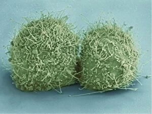
Background imageScanning Electron Micrograph Collection: Scanning electron micrograph of just-divided HeLa cells

Scanning electron micrograph of just-divided HeLa cells. Zeiss Merlin HR-SEM

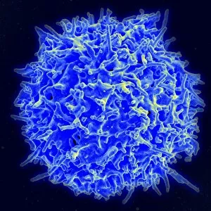
Background imageScanning Electron Micrograph Collection: Scanning electron micrograph of a human T cell

Scanning electron micrograph of a human T cell
Scanning electron micrograph of a human T lymphocyte (also called a T cell) from the immune system of a healthy donor

Background imageScanning Electron Micrograph Collection: Colorized scanning electron micrograph of a T lymphocyte

Colorized scanning electron micrograph of a T lymphocyte

Background imageScanning Electron Micrograph Collection: Scanning electron micrograph of a schistosome parasite

Scanning electron micrograph of a schistosome parasite, which enters the body through the skin of persons coming in contact with infested waters. The adult worm lives in the veins of its host

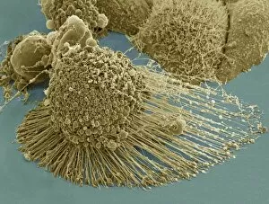
Background imageScanning Electron Micrograph Collection: Scanning electron micrograph of an apoptotic HeLa cell

Scanning electron micrograph of an apoptotic HeLa cell. Zeiss Merlin HR-SEM

Background imageScanning Electron Micrograph Collection: Papilio palinurus, emerald swallowtail

Papilio palinurus, emerald swallowtail
SEM image of an emerald swallowtails wing

Background imageScanning Electron Micrograph Collection: Pieris rapae, small white

Pieris rapae, small white
SEM image of the wing of a small white butterfly

Background imageScanning Electron Micrograph Collection: Papilio machaon, old world swallowtail

Papilio machaon, old world swallowtail
SEM image of Papilio machaon wing

Background imageScanning Electron Micrograph Collection: Papilio palinurus, emerald swallowtail

Papilio palinurus, emerald swallowtail
SEM image of an emerald swallowtails wing

Background imageScanning Electron Micrograph Collection: Malachite

Malachite comprises of (copper carbonate hydroxide). Malachite has distinctive green banding and belongs to the carbonate class

Background imageScanning Electron Micrograph Collection: Scyphosphaera apsteinii

Scyphosphaera apsteinii. SEM image of an equatorial coccolith

Background imageScanning Electron Micrograph Collection: Dinosaur eggshell

Dinosaur eggshell
Scanning electron microscope image on display in the Darwin Centre

Background imageScanning Electron Micrograph Collection: Ventral surface of a mite from the prostigmatic species

Ventral surface of a mite from the prostigmatic species
Scanning electron microscope image displayed on the glass screens in the Darwin Centre, at the Natural History Museum, London

Background imageScanning Electron Micrograph Collection: Visbyshaera oligofurcata, acritarch

Visbyshaera oligofurcata, acritarch
Scanning electron microscope image of a microfossil belonging to a group of marine phytoplanktonic organisms known as acritarchs that teemed in Silurian seas about 415 Ma ago


All Professionally Made to Order for Quick Shipping

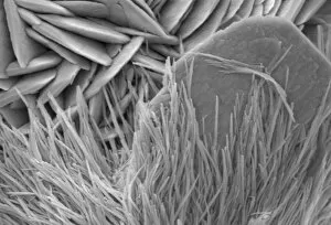
"Exploring the Microscopic World: Captivating Scanning Electron Micrographs" Witness the intricate beauty of nature with this Scanning Electron Micrograph (SEM) capturing a Praying Mantis in stunning detail, magnified 30 times its size. Delve into the microscopic realm and discover the resilience of Tardigrades, also known as 'Water Bears, ' through this SEM image magnified at an astounding 1250 times. Uncover the hidden dangers lurking within Crysotile asbestos as revealed by this powerful SEM, shedding light on its harmful effects on human health. Journey inside the liver through a fascinating SEM, offering a glimpse into its complex structure and vital role in our body's functions. Get up close and personal with Cimex lectularius, commonly known as bed bugs, thanks to this detailed SEM that exposes their tiny yet formidable presence. Marvel at the intricate features of a Fruit Fly captured under an SEM lens at 300 times magnification – revealing astonishing details invisible to the naked eye. Explore Taraxacum officinale or dandelion's fruiting head like never before with this captivating SEM image showcasing its delicate structures and unique design. Discover nature's engineering marvels by examining Snail teeth under high-powered microscopy; witness how these minuscule structures aid them in feeding and survival. Peer into another world within Plasmodium sp. , a malarial parasite responsible for widespread disease transmission worldwide – all unveiled through this striking SEM image. Dive deep into geology with Kaolinite - explore its fine crystalline structure using an advanced scanning electron microscope to unravel its mineralogical secrets. Encounter Sarcoptes scabiei or scabies mite up close via an extraordinary SEM capture; gain insight into their morphology while appreciating their impact on human health.