Fraud Blocker Skip to main content

Scanner Collection (page 4)

"Unveiling the Hidden Wonders: The Scanner's Journey" Delicate and ethereal

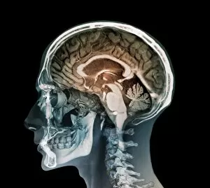
Background imageScanner Collection: Human head, 3D CT scan C016 / 6377

Human head, 3D CT scan C016 / 6377
Human head. Coloured 3D computed tomography (CT) scan of a profile view of a 47 year old patients head. The brain is purple and bone is light brown. At centre, behind the face, are the sinuses

Background imageScanner Collection: Stag beetle C016 / 2181

Stag beetle C016 / 2181
Stag beetle

Background imageScanner Collection: Leaf insect C016 / 2231

Leaf insect C016 / 2231
Leaf insect. View of the upperside of a Linnaeus leaf insect (Phyllium siccifolium). Specimen obtained from the University of Valencia, Spain

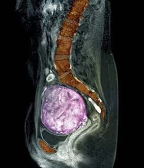
Background imageScanner Collection: Uterine fibroids, MRI scan C018 / 0467

Uterine fibroids, MRI scan C018 / 0467
Uterine fibroids. Coloured magnetic resonance imaging (MRI) scan of the pelvis of a 47 year old female with two large fibroids (pink) in her uterus

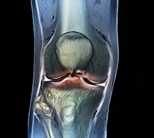
Background imageScanner Collection: Healthy knee, CT scan C018 / 0415

Healthy knee, CT scan C018 / 0415
Healthy knee. Coloured frontal computed tomography (CT) scan projected on to a magnetic resonance imaging (MRI) scan of the knee of a 30 year old

Background imageScanner Collection: Rectal cancer, MRI scans C018 / 0508

Rectal cancer, MRI scans C018 / 0508
Rectal cancer. Coloured magnetic resonance imaging (MRI) scans of the pelvis of a 65 year old patient with a malignant (cancerous) tumour (purple) in the rectum, the last part of the large intestine
Background imageScanner Collection: Rectal cancer, MRI scans C018 / 0510

Rectal cancer, MRI scans C018 / 0510
Rectal cancer. Coloured magnetic resonance imaging (MRI) scans of the pelvis of a 65 year old patient with a malignant (cancerous) tumour in the rectum, the last part of the large intestine

Background imageScanner Collection: Spinal cord cyst, MRI scan C018 / 0538

Spinal cord cyst, MRI scan C018 / 0538
Spinal cord cyst. Magnetic resonance imaging (MRI) scan of the cervical (neck) spinal cord of a 28 year old patient with syringomyelia, a cyst in the spinal cord. A cyst is a fluid-filled sac

Background imageScanner Collection: Healthy knee, CT scan C018 / 0414

Healthy knee, CT scan C018 / 0414
Healthy knee. Coloured frontal computed tomography (CT) scan projected on to a magnetic resonance imaging (MRI) scan of the knee of a 30 year old

Background imageScanner Collection: Healthy lumbar spine, MRI C018 / 0563

Healthy lumbar spine, MRI C018 / 0563
Healthy lumbar spine. Coloured magnetic resonance imaging (MRI) scan through the disc (red circle) between the second and third lumbar (lower back) vertebrae of a 30 year old patient

Background imageScanner Collection: Healthy brain, MRI scan C018 / 0431

Healthy brain, MRI scan C018 / 0431
Healthy brain. Coloured 3D magnetic resonance imaging (MRI) scan of the healthy brain (excluding the brainstem) of a 37 year old. The cavities are the ventricles

Background imageScanner Collection: Healthy brain, MRI scan C018 / 0430

Healthy brain, MRI scan C018 / 0430
Healthy brain. Coloured 3D magnetic resonance imaging (MRI) scan of the healthy brain (excluding the brainstem) of a 37 year old. The cavities are the ventricles

Background imageScanner Collection: Uterine fibroids, MRI scan C018 / 0468

Uterine fibroids, MRI scan C018 / 0468
Uterine fibroids. Coloured magnetic resonance imaging (MRI) scan of the pelvis of a 47 year old female with two large fibroids (pink) in her uterus

Background imageScanner Collection: Healthy brain, MRI scan C018 / 0429

Healthy brain, MRI scan C018 / 0429
Healthy brain. Coloured 3D magnetic resonance imaging (MRI) scan of the healthy brain (excluding the brainstem) of a 37 year old. The cavities are the ventricles

Background imageScanner Collection: Cerebrotendinous xanthomatosis, MRI scan C018 / 0504

Cerebrotendinous xanthomatosis, MRI scan C018 / 0504
Cerebrotendinous xanthomatosis. Coloured magnetic resonance imaging (MRI) scan of the brain of a 46 year old patient with cerebrotendinous xanthomatosis (CTX)

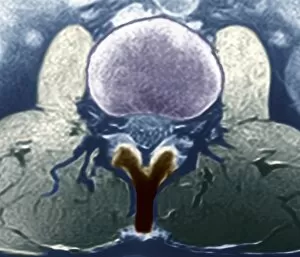
Background imageScanner Collection: Healthy lumbar spine, MRI C018 / 0562

Healthy lumbar spine, MRI C018 / 0562
Healthy lumbar spine. Coloured magnetic resonance imaging (MRI) scan through the disc (purple) between the second and third lumbar (lower back) vertebrae of a 30 year old patient

Background imageScanner Collection: Rectal cancer, MRI scans C018 / 0511

Rectal cancer, MRI scans C018 / 0511
Rectal cancer. Coloured magnetic resonance imaging (MRI) scans of the pelvis of a 65 year old patient with a malignant (cancerous) tumour (purple) in the rectum, the last part of the large intestine

Background imageScanner Collection: Uterine fibroids, MRI scan C018 / 0469

Uterine fibroids, MRI scan C018 / 0469
Uterine fibroids. Coloured magnetic resonance imaging (MRI) scan of the pelvis of a 47 year old female with two large fibroids (pink) in her uterus

Background imageScanner Collection: Healthy abdomen, MRI scan C018 / 0663

Healthy abdomen, MRI scan C018 / 0663
Healthy abdomen. Coloured magnetic resonance imaging (MRI) scan of the abdomen of a 35 year old patient showing the internal organs. At top are the stomach (purple) and liver (red)

Background imageScanner Collection: Healthy abdomen, MRI scan C018 / 0664

Healthy abdomen, MRI scan C018 / 0664
Healthy abdomen. Coloured magnetic resonance imaging (MRI) scan of the abdomen of a 35 year old patient showing the internal organs. At top are the stomach (brown) and liver (orange)

Background imageScanner Collection: Early-onset Alzheimers disease, MRI scan

Early-onset Alzheimers disease, MRI scan
Early-onset Alzheimers disease. Magnetic resonance imaging (MRI) scans of the brain of a 53 year old patient with early-onset Alzheimers disease

Background imageScanner Collection: Rectal cancer, MRI scan C018 / 0512

Rectal cancer, MRI scan C018 / 0512
Rectal cancer. Coloured magnetic resonance imaging (MRI) scan of the pelvis of a 65 year old patient with a malignant (cancerous) tumour in the rectum, the last part of the large intestine

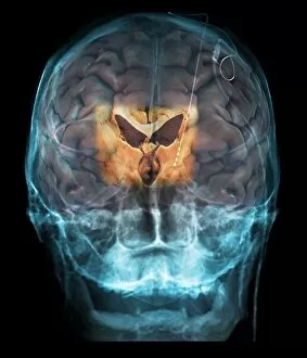
Background imageScanner Collection: Brain implants for Parkinsons disease

Brain implants for Parkinsons disease. Coloured 3D magnetic resonance imaging (MRI) scan of the brain of a patient who has had an electrode (white line) inserted into the brain

Background imageScanner Collection: Healthy brain, MRI scan C018 / 0423

Healthy brain, MRI scan C018 / 0423
Healthy brain. Coloured 3D magnetic resonance imaging (MRI) scan of the healthy brain (excluding the brainstem) of a 37 year old. The cavities are the ventricles

Background imageScanner Collection: Human head, MRI and 3D CT scans C018 / 0636

Human head, MRI and 3D CT scans C018 / 0636
Human head. Coloured composite image of a magnetic resonance imaging (MRI) scan of the brain and 2D and 3D computed tomography (CT) scans of the head and neck of a 35 year old patient

Background imageScanner Collection: Spinal cord cyst, MRI scan C018 / 0561

Spinal cord cyst, MRI scan C018 / 0561
Spinal cord cyst. Coloured magnetic resonance imaging (MRI) scan of the cervical (neck) spinal cord of a 28 year old patient with syringomyelia, a cyst in the spinal cord

Background imageScanner Collection: Genetic analysis, scanning samples C019 / 0258

Genetic analysis, scanning samples C019 / 0258
Genetic analysis. Analyst in a genetics laboratory, scanning labelled blood samples. DNA (deoxyribonucleic acid) will be isolated from the samples

Background imageScanner Collection: Scanning genetic microarray C019 / 0285

Scanning genetic microarray C019 / 0285
Scanning genetic microarray. Analyst using a scanner to read the barcode of a gene chip or microarray. Samples of DNA (deoxyribonucleic acid) have been added to the microarray

Background imageScanner Collection: Scanning genetic microarray C019 / 0286

Scanning genetic microarray C019 / 0286
Scanning genetic microarray. Analyst using a scanner to read the barcode of a gene chip or microarray. Samples of DNA (deoxyribonucleic acid) have been added to the microarray

Background imageScanner Collection: Digital camera sensitometry C014 / 2085

Digital camera sensitometry C014 / 2085
The imaging characteristics of a digital camera can be assessed by using the sensitometric methods developed for silver-based photographic systems

Background imageScanner Collection: Human torso, coloured mri scan C014 / 2644

Human torso, coloured mri scan C014 / 2644
Normal human torso, coloured frontal magnetic resonance imaging (MRI) scan. The spinal bones (vertebrae) are seen running from the top of the chest to the pelvis

Background imageScanner Collection: Alcohol detector for traffic police

Alcohol detector for traffic police
Drunk driving. A laser-based detector that can remotely sense alcohol fumes inside a passing car on show. The device is aimed at allowing traffic police to detect drivers who have had alcohol by

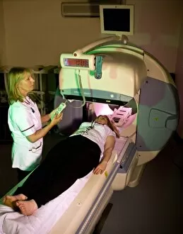
Background imageScanner Collection: Medical imaging in lung disease research

Medical imaging in lung disease research. Radiologist preparing a gamma camera and computed tomography (CT) scanner to produce 3D images of a patients lungs

Background imageScanner Collection: Fingerprint scan, conceptual image C013 / 5597

Fingerprint scan, conceptual image C013 / 5597
Fingerprint scan, conceptual image

Background imageScanner Collection: Brain injury, MRI scan

Brain injury, MRI scan
Brain injury. Coronal magnetic resonance imaging (MRI) brain scan showing intracranial abnormalities in a 23-year-old male motorcycle crash victim

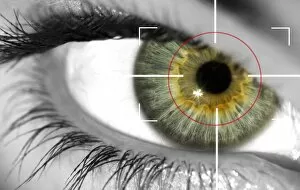
Background imageScanner Collection: Biometrics, conceptual image

Biometrics, conceptual image
MODEL RELEASED. Biometrics, conceptual composite image

Background imageScanner Collection: Anthophora hispanica bee C016 / 2081

Anthophora hispanica bee C016 / 2081
Anthophora hispanica bee

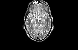
Background imageScanner Collection: MRI scan

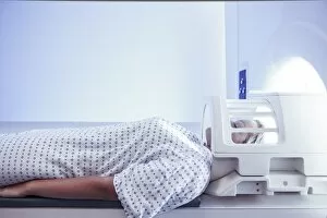
MRI scan

Background imageScanner Collection: Fractured pelvis, CT scan C016 / 6584

Fractured pelvis, CT scan C016 / 6584
Fractured pelvis. Coloured computed tomography (CT) scan of the pelvis of a 56 year old that was in a road traffic accident

Background imageScanner Collection: Pulmonary embolism, CT scan

Pulmonary embolism, CT scan
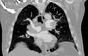
Pulmonary embolism. Computed tomography (CT) scan of a section through a 65 year old male patients chest showing blood clots, or embolisms (white, centre left adn centre right)

Background imageScanner Collection: Mesothelioma lung cancer, CT scan

Mesothelioma lung cancer, CT scan
Mesothelioma lung cancer. Computed tomography (CT) scan of the lungs of a 69 year old patient with mesothelioma, a type of lung cancer

Background imageScanner Collection: Lung cancer, CT and PET scans

Lung cancer, CT and PET scans
Lung cancer. Combined computed tomography (CT) and positron emission tomography (PET) scans of the lungs of an 84 year old woman with a malignant (cancerous) tumour (bright patch)

Background imageScanner Collection: Polycystic kidneys, ultrasound scan C017 / 7747

Polycystic kidneys, ultrasound scan C017 / 7747
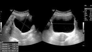
Polycystic kidneys. Ultrasound scan of the kidneys of a 42 year old female showing numerous fluid-filled cysts (dark areas)

Background imageScanner Collection: Brain cancer, MRI scan C016 / 4438

Brain cancer, MRI scan C016 / 4438
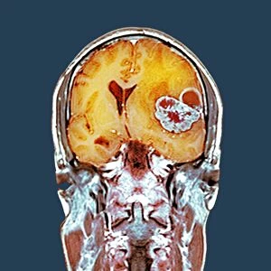
Brain cancer. Coloured coronal magnetic resonance imaging (MRI) scan through the head of a 48 year old male patient with a glioblastoma (red)

Background imageScanner Collection: Brain cancer, MRI scan C016 / 4436

Brain cancer, MRI scan C016 / 4436
Brain cancer. Coloured coronal magnetic resonance imaging (MRI) scan through the head of a 48 year old male patient with a glioblastoma (centre right)

Background imageScanner Collection: Brain cancer, MRI scan C016 / 4437

Brain cancer, MRI scan C016 / 4437
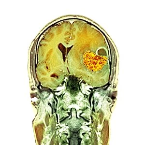
Brain cancer. Coloured coronal magnetic resonance imaging (MRI) scan through the head of a 48 year old male patient with a glioblastoma (orange)

Background imageScanner Collection: QR code after being scanned C013 / 9472

QR code after being scanned C013 / 9472
QR code (on bottle) after being scanned by a mobile phone. A QR code, designed to be scanned by devices such as mobile phones

Background imageScanner Collection: QR code being scanned C013 / 9473

QR code being scanned C013 / 9473
QR code (lower right) being scanned by a tablet computer. A QR code, designed to be scanned by devices such as mobile phones or mobile-enabled computers


All Professionally Made to Order for Quick Shipping

"Unveiling the Hidden Wonders: The Scanner's Journey" Delicate and ethereal, a sea green swallowtail butterfly gracefully flutters its wings under the watchful eyes of a scanner. With precision and accuracy, a full body scan captures every detail, revealing the intricate workings of our complex human form. Peering into the depths of brain anatomy, an MRI scan unveils the mysteries hidden within, unlocking secrets that shape our thoughts and actions. Like a unique signature, a fingerprint scanner maps out individual patterns with meticulous care, ensuring security in an ever-evolving world. Injured but not defeated, an MRI C018 / 0649 reveals the extent of damage to a ruptured Achilles tendon - guiding doctors towards effective treatment options. Beyond fingerprints' utilitarian purpose lies artistic beauty; an artwork created by capturing each swirl through a fingerprint scanner tells tales untold before. A glimpse into normalcy is found as an MRI C016 / 8845 showcases the intricacies of a healthy human brain - reminding us of its remarkable resilience. Seeking answers beneath layers unseen, an MRI exposes ruptured breast implants - providing clarity for those seeking resolution and healing. Innocence encapsulated within imaging technology as we witness childlike wonder through glimpses into young minds via captivating MRI scans. Unraveling connections that make us who we are; white matter fibers come alive under DTI scans - illuminating pathways crucial for communication within our brains. Echoing familiarity amidst diversity; another snapshot from everyday life emerges with Normal Human Brain captured by MRI C016 / 8850 – reminding us how extraordinary ordinary can be.