
Scan Collection (page 7)
"Unveiling the Unseen: The Power of Scanning" Exploring the Past: The Old Hartlepool Lighthouse stands tall as a beacon of history in north east England


All Professionally Made to Order for Quick Shipping
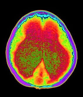
"Unveiling the Unseen: The Power of Scanning" Exploring the Past: The Old Hartlepool Lighthouse stands tall as a beacon of history in north east England. Peering Inside: Discovering the Hidden Truths through Full Body Scans and MRI scans. Battling Brain Tumors: How MRI scans aid in early detection and treatment of brain tumors. Art Meets Technology: Fingerprint scanners bring security to life while enhancing artwork's allure. Decoding Secrets: DDE-90036800 reveals an intriguing mystery waiting to be unraveled. Illuminating Minds: Illustrating the intricacies of a normal brain through captivating MRI scans. Fighting Back Against Meningitis: How MRI scans play a crucial role in diagnosing bacterial meningitis. Security Check Revealed: A woman walks confidently through a security gate, her bag's contents made visible by scanning technology. Visualizing Hope Amidst Challenges: 3-D MRI scans aiding doctors' understanding and treatment of brain tumors. Artistic Wonders within Our Brains: Captivating artworks inspired by mesmerizing brain scan imagery. Modernizing Safety Measures at Aircraft Depots with X-ray Scans for cadets - DDE-90039098 showcases progress. In this ever-evolving world, scanning technologies have become our window into hidden realms – from unraveling historical mysteries at landmarks like the Old Hartlepool Lighthouse, to peering inside our bodies using full body and MRI scans that hold potential lifesaving information about conditions such as brain tumors or bacterial meningitis. Beyond medical applications, fingerprint scanners merge artistry with security measures, creating visually stunning masterpieces while ensuring safety is not compromised. The enigmatic code "DDE-90036800" hints at untold stories waiting to be deciphered; it holds secrets yet unknown, inviting us to embark on a thrilling journey of discovery.