
Reproductive Structure Collection
"Exploring the Intricate World of Reproductive Structures: From Pollen Grains to Ovarian Follicles" Delving into the microscopic realm


All Professionally Made to Order for Quick Shipping
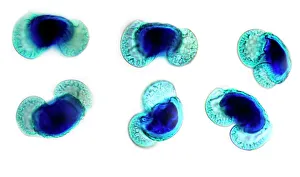
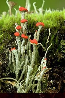
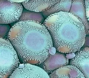
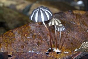
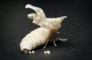
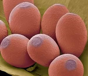
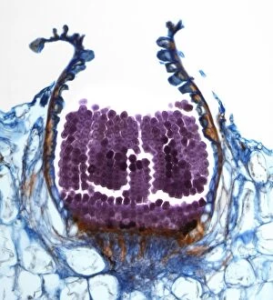
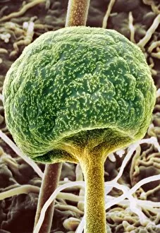
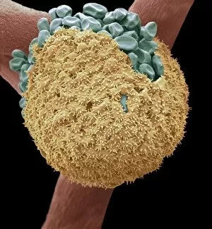
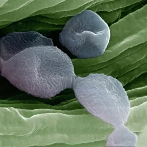
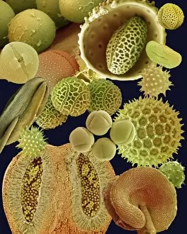
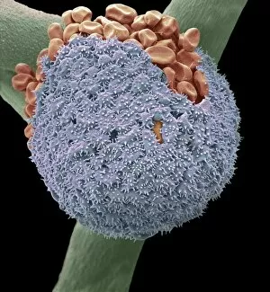
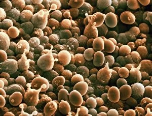
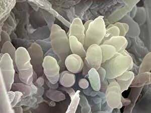
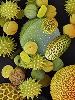
"Exploring the Intricate World of Reproductive Structures: From Pollen Grains to Ovarian Follicles" Delving into the microscopic realm, SEM reveals the stunning beauty of pollen grains, showcasing their intricate structures and diverse shapes. Pine pollen grains captured under a light micrograph unveil their unique features, highlighting nature's remarkable attention to detail in reproductive mechanisms. The cup lichen (Cladonia floerkeana) exhibits its reproductive prowess through captivating textures and patterns, demonstrating how life finds a way even in harsh environments. A closer look at an ovarian follicle through a light micrograph (C016 / 0519) unveils the delicate process of female reproduction with awe-inspiring precision. Zooming in on a common daisy anther using SEM exposes its fascinating surface details, shedding light on how plants ensure successful pollination for future generations. An orange fruit captured under a light micrograph showcases the culmination of successful reproduction – vibrant colors and enticing textures that entice animals to disperse seeds far and wide. Brassica pollen viewed through SEM provides insight into this plant family's unique reproductive strategies, unraveling secrets hidden within tiny grains that hold immense potential for new life. Thale cress flower depicted in a mesmerizing micrograph exemplifies nature's artistry as it orchestrates complex processes leading to fertilization and seed production. Bread mould examined via SEM unravels its intriguing spore-bearing structures, reminding us that even seemingly insignificant organisms play crucial roles in ecosystems' reproductive cycles. Rust fungus infection magnified by SEM offers both scientific intrigue and cautionary tales about how pathogens exploit host plants' reproductive systems for their own propagation. Broad bean pollen observed under SEM lenses presents an array of sculpted forms designed for efficient transfer between flowers - ensuring genetic diversity within populations.