
Radiological Collection
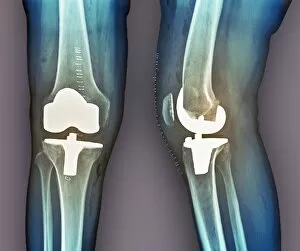
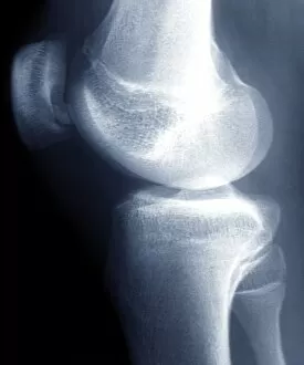
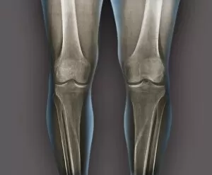
"Unlocking the Secrets: Radiological Insights into Our Bodies" Revealing a Healthy Knee


All Professionally Made to Order for Quick Shipping
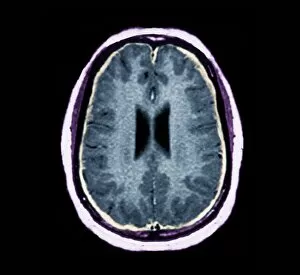
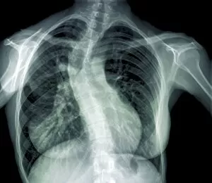
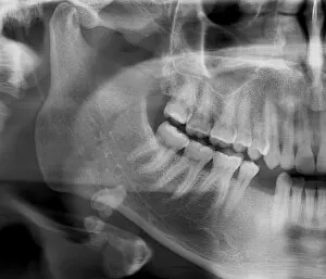
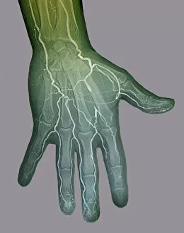
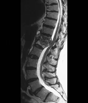
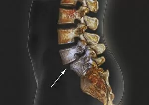
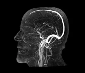
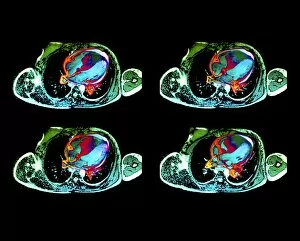
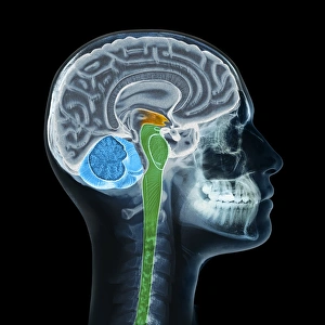
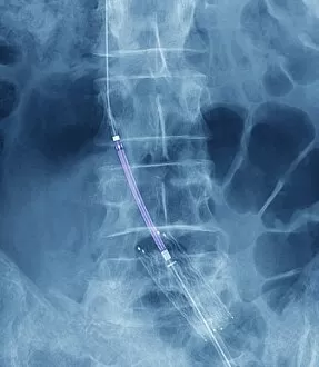
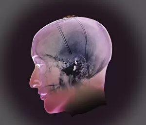
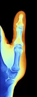
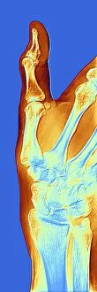
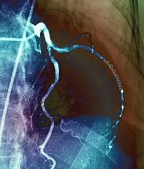
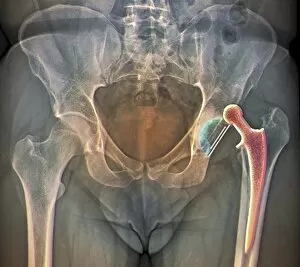
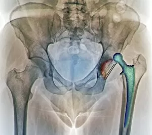
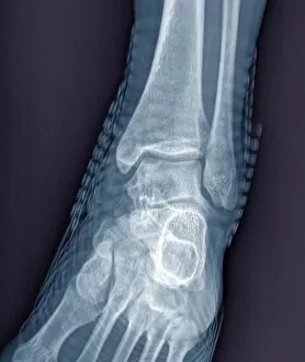
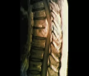
"Unlocking the Secrets: Radiological Insights into Our Bodies" Revealing a Healthy Knee: A closer look through X-ray imaging showcases the strength and resilience of a healthy knee joint. Unmasking Bacterial Meningitis: An MRI scan provides vital radiological evidence in diagnosing bacterial meningitis, aiding doctors in timely treatment. On-the-Go Imaging Solutions: The advent of mobile x-ray units brings convenience and efficiency to healthcare, ensuring prompt diagnosis and care wherever needed. Restoring Mobility with Total Knee Replacement: X-rays play a crucial role in assessing successful outcomes after total knee replacement surgeries, offering insights into improved mobility for patients. Peering into a Healthy Ankle Joint: Through an x-ray lens, witness the intricate structure of a healthy ankle joint that supports our daily activities without hindrance. Detecting Scoliosis through X-ray Vision: X-rays help identify scoliosis early on, enabling effective treatment plans to alleviate discomfort and maintain spinal health. Mapping Normalcy with Leg X-rays: Explore the fascinating skeletal framework of normal legs captured by x-ray technology – an invaluable tool for orthopedic assessments. Visualizing Ischaemia's Impact with Digital Angiograms: Digital angiograms provide detailed imagery to evaluate blood flow disruptions caused by ischaemia, guiding targeted interventions for better patient outcomes. Decoding Arthrosis of the Hand via X-Ray F006/4616: Delve into the intricacies of arthrosis affecting hand joints using specialized x-rays (F006/4616), aiding accurate diagnoses and personalized treatments. Illuminating Brain Aneurysms through 3D Scans: Cutting-edge 3D scanning techniques shed light on brain aneurysms' complex anatomy, assisting neurosurgeons in planning precise interventions for optimal patient care. Spotting Tuberculosis of the Spine with MRI Scans.