Fraud Blocker Skip to main content

Radiography Collection (page 3)

Radiography, the art of capturing hidden wonders through the lens of X-rays, unveils a captivating world beyond our naked eyes

Background imageRadiography Collection: Michaelmas daisy (Aster amellus) flower head, X-ray

Michaelmas daisy (Aster amellus) flower head, X-ray

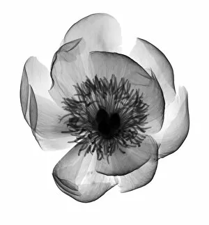
Background imageRadiography Collection: Peony (Paeonia sp. ), X-ray

Peony (Paeonia sp. ), X-ray
Peony (Paeonia sp.), X-ray

Background imageRadiography Collection: Lotus flower (Nelumbo nucifera), X-ray

Lotus flower (Nelumbo nucifera), X-ray

Background imageRadiography Collection: Magnolia flower and acai berries, X-ray

Magnolia flower and acai berries, X-ray

Background imageRadiography Collection: Gardenia head from above, X-ray

Gardenia head from above, X-ray
Gardenia sp. head from above, X-ray

Background imageRadiography Collection: Osteoarthritis of the knee, X-ray

Osteoarthritis of the knee, X-ray
Osteoarthritis of the knee, coloured X-ray

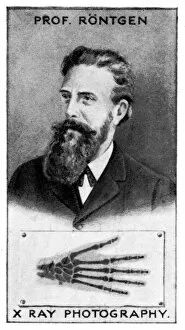
Background imageRadiography Collection: Wilhelm Conrad Roentgen, German physicist, (c1924)

Wilhelm Conrad Roentgen, German physicist, (c1924). On 8 November 1895, Roentgen (or Rontgen, 1845-1923) discovered what came to be known as X-rays

Background imageRadiography Collection: Radiography controls JLP01_08_069288

Radiography controls JLP01_08_069288
West Cumberland Hospital, Homewood Road, Homewood, Whitehaven, Copeland, Cumbria. An x-ray room in the radiography department at West Cumberland Hospital showing the control booth

Background imageRadiography Collection: Radiography JLP01_10_34515

Radiography JLP01_10_34515
St Georges Hospital, Blackshaw Road, Tooting, Wandsworth, Greater London. Two radiographers adjusting x-ray equipment at St Georges Hospital, Tooting

Background imageRadiography Collection: Flowers in vase, X-ray

Flowers in vase, X-ray
Flowers in vase, coloured X-ray

Background imageRadiography Collection: Violet (Viola odorata) leaf in vase, X-ray

Violet (Viola odorata) leaf in vase, X-ray
Violet (Viola odorata) leaf in vase, coloured X-ray

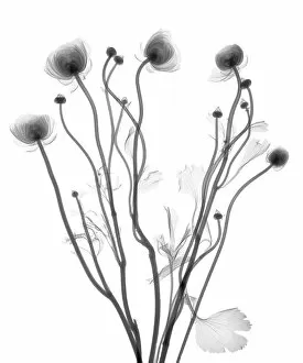
Background imageRadiography Collection: Buttercup (Ranunculus sp. ), X-ray

Buttercup (Ranunculus sp. ), X-ray
Buttercup (Ranunculus sp.), X-ray

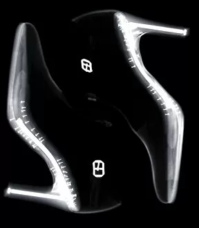
Background imageRadiography Collection: High-heels, X-ray

High-heels, X-ray

Background imageRadiography Collection: High heel shoe, X-ray

High heel shoe, X-ray

Background imageRadiography Collection: High heel shoes, X-ray

High heel shoes, X-ray

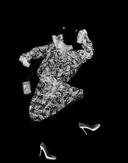
Background imageRadiography Collection: Dancing dress, shoes and MP3, X-ray

Dancing dress, shoes and MP3, X-ray

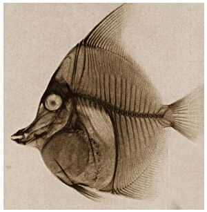
Background imageRadiography Collection: X Ray of a fish c. 1890

X Ray of a fish c. 1890
3501927 X Ray of a fish c.1890 by German School, (19th century); Private Collection; Prismatic Pictures; German, out of copyright

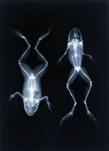
Background imageRadiography Collection: An early X-ray photo of frogs by Joseph Maria Eder. 1896 (photogravure)

An early X-ray photo of frogs by Joseph Maria Eder. 1896 (photogravure)
746718 An early X-ray photo of frogs by Joseph Maria Eder. 1896 (photogravure) by Austrian School, (19th century); Private Collection; (add.info)

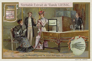
Background imageRadiography Collection: Radiography and X-rays (chromolitho)

Radiography and X-rays (chromolitho)
974602 Radiography and X-rays (chromolitho) by European School, (19th century); Private Collection; (add.info.: Radiography and X-rays)

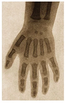
Background imageRadiography Collection: X Ray of a childs hand c. 1890

X Ray of a childs hand c. 1890
3501926 X Ray of a childs hand c.1890 by German School, (19th century); Private Collection; Prismatic Pictures; German, out of copyright

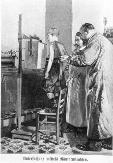
Background imageRadiography Collection: Wilhelm Konrad Roentgen (1845-1923) x-raying a young boy, from a book by Hans Kraemer, c

Wilhelm Konrad Roentgen (1845-1923) x-raying a young boy, from a book by Hans Kraemer, c
CHT237292 Wilhelm Konrad Roentgen (1845-1923) x-raying a young boy, from a book by Hans Kraemer, c.1898-99 (b/w photo) by German School, (19th century); Bibliotheque Nationale, Paris

Background imageRadiography Collection: X Ray of an ankle c. 1890

X Ray of an ankle c. 1890
3501925 X Ray of an ankle c.1890 by German School, (19th century); Private Collection; Prismatic Pictures; German, out of copyright

Background imageRadiography Collection: Radiguet x-ray machine (engraving) (b / w photo)

Radiguet x-ray machine (engraving) (b / w photo)
CHT237293 Radiguet x-ray machine (engraving) (b/w photo) by French School, (19th century); Bibliotheque Nationale, Paris, France; eArchives Charmet; French, out of copyright

Background imageRadiography Collection: X Ray of a fractured wrist c. 1890

X Ray of a fractured wrist c. 1890
3501929 X Ray of a fractured wrist c.1890 by German School, (19th century); Private Collection; Prismatic Pictures; German, out of copyright

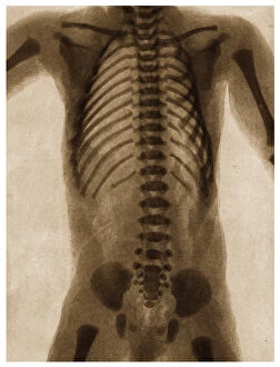
Background imageRadiography Collection: X Ray of a human torso c. 1890

X Ray of a human torso c. 1890
3501928 X Ray of a human torso c.1890 by German School, (19th century); Private Collection; Prismatic Pictures; German, out of copyright

Background imageRadiography Collection: Inside an early x-ray room (b / w photo)

Inside an early x-ray room (b / w photo)
STC343848 Inside an early x-ray room (b/w photo) by German Photographer, (19th century); Private Collection; (add.info.: X-rays were discovered by Wilhelm Conrad RAontgen (1845-1923) in 1895

Background imageRadiography Collection: The mass X-ray campaign for tuberculosis, Liverpool. Pictured, Mrs Bessie Braddock

The mass X-ray campaign for tuberculosis, Liverpool. Pictured, Mrs Bessie Braddock, MP for Exchange Division of Liverpool, being greeted by Dr Andrew B Semple, Liverpools Medical Officer of Health

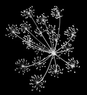
Background imageRadiography Collection: Cow parsley (Petroselinum crispum), X-ray

Cow parsley (Petroselinum crispum), X-ray

Background imageRadiography Collection: Honeycomb, X-ray

Honeycomb, X-ray

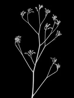
Background imageRadiography Collection: Kangaroo paw (Anigozanthos sp. ), X-ray

Kangaroo paw (Anigozanthos sp. ), X-ray
Kangaroo paw (Anigozanthos sp.), X-ray

Background imageRadiography Collection: Two tulips, X-ray

Two tulips, X-ray

Background imageRadiography Collection: Two alliums, X-ray

Two alliums, X-ray

Background imageRadiography Collection: Mountain ash (Sorbus rosaceae), X-ray

Mountain ash (Sorbus rosaceae), X-ray

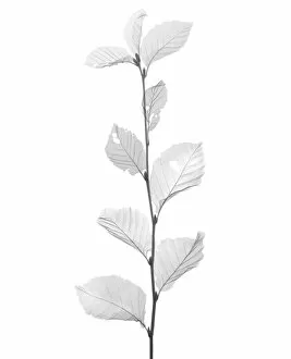
Background imageRadiography Collection: Beech leaves (Fagus sp. ), X-ray

Beech leaves (Fagus sp. ), X-ray
Beech leaves (Fagus sp.), X-ray

Background imageRadiography Collection: Two green fig (Ficus carica) leaves overlapping, X-ray

Two green fig (Ficus carica) leaves overlapping, X-ray
Two green fig (Ficus carica) leaves overlapping, coloured X-ray

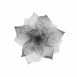
Background imageRadiography Collection: Wild flower (Anemone sp. ), X-ray

Wild flower (Anemone sp. ), X-ray
Wild flower (Anemone sp.), X-ray

Background imageRadiography Collection: Camellia, X-ray

Camellia, X-ray

Background imageRadiography Collection: Fern (Davallia mariessii), X-ray

Fern (Davallia mariessii), X-ray

Background imageRadiography Collection: Dried lisianthus (Eustoma grandiflorum), X-ray

Dried lisianthus (Eustoma grandiflorum), X-ray

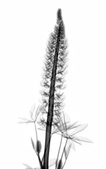
Background imageRadiography Collection: Lupin (Lupinus sp. ), X-ray

Lupin (Lupinus sp. ), X-ray
Lupin (Lupinus sp.), X-ray

Background imageRadiography Collection: Maple (Acer sp. ), X-ray

Maple (Acer sp. ), X-ray
Maple (Acer sp.), X-ray

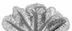
Background imageRadiography Collection: Crest cabbage (Brassica sp. ), X-ray

Crest cabbage (Brassica sp. ), X-ray
Crest cabbage (Brassica sp.), X-ray

Background imageRadiography Collection: Two branches with leaves, X-ray

Two branches with leaves, X-ray

Background imageRadiography Collection: Festive lights in shape of a tree, X-ray

Festive lights in shape of a tree, X-ray
Festive lights in shape of a tree, coloured X-ray

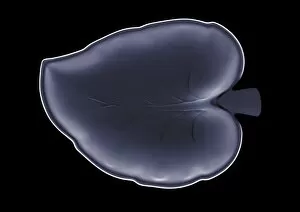
Background imageRadiography Collection: Leaf plate, X-ray

Leaf plate, X-ray

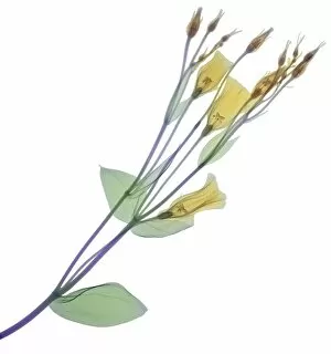
Background imageRadiography Collection: Branch with multiple yellow flowers and buds, X-ray

Branch with multiple yellow flowers and buds, X-ray
Branch with multiple yellow flowers and buds, coloured X-ray

Background imageRadiography Collection: Pink flower with leaves, X-ray

Pink flower with leaves, X-ray
Pink flower with leaves, coloured X-ray

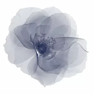
Background imageRadiography Collection: Pastel blue lily (Anthurium sp. ), X-ray

Pastel blue lily (Anthurium sp. ), X-ray
Pastel blue lily (Anthurium sp.), coloured X-ray


All Professionally Made to Order for Quick Shipping

Radiography, the art of capturing hidden wonders through the lens of X-rays, unveils a captivating world beyond our naked eyes. Like the awe-inspiring "Pillars of Creation" in space, radiography allows us to explore intricate details within our own bodies and beyond. Delving into the depths of human anatomy, it reveals the delicate network of brain blood vessels with a mesmerizing 3D angiogram from 1981. A broken wrist bone is laid bare by an X-ray, showcasing both fragility and resilience in one image. Beyond humans, radiography takes us on a journey through nature's marvels. The majestic skull of a horse reminds us of their grace and strength. Meanwhile, even skeletons find joy as we witness one playing rugby - an unexpected twist that brings humor to this scientific art form. But radiography doesn't stop at living beings; it delves into dental health too. A panoramic dental X-ray captures every tooth's story while revealing potential issues lurking beneath the surface. Even marine life isn't spared from its gaze: an ethereal lobster appears ghostly yet beautiful when seen through an X-ray lens. The fusion between technology and humanity becomes evident as we encounter a person holding a camera captured in an X-ray frame – reminding us that even those behind the lens are not exempt from exploration. Intriguingly surreal images emerge as skeletons engage in activities typically associated with life: drinking or engaging in sports like rugby. These playful depictions remind us that there is always more than meets the eye when it comes to our inner structures. Radiography transcends earthly boundaries too; it ventures into outer space where composite images reveal spiral galaxy M81's cosmic dance among stars – merging science and art seamlessly together. From triggerfish to seahorses, radiography exposes intricate skeletal structures unseen under normal circumstances—reminding us that beauty exists even within what may seem ordinary or overlooked.