
Radiographer Collection
"Unveiling the Hidden: A Journey into the World of Radiographers" Step back in time to witness the birth of radiography, as captured through captivating X-ray images


All Professionally Made to Order for Quick Shipping
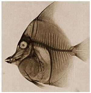
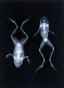
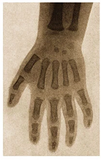
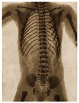
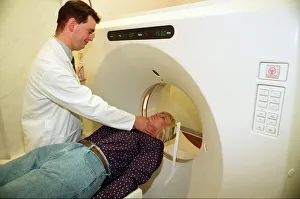
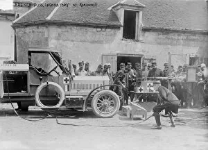
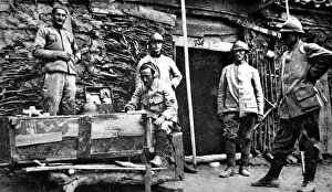
"Unveiling the Hidden: A Journey into the World of Radiographers" Step back in time to witness the birth of radiography, as captured through captivating X-ray images. Delve into history with an intriguing X-ray of a fish from 1890, revealing its intricate skeletal structure like never before. Marvel at Joseph Maria Eder's groundbreaking early X-ray photo of frogs in 1896, immortalized through photogravure. The power of radiography extends beyond nature's realm; explore the delicate innocence encapsulated within an X-ray image of a child's hand from c. 1890. Witness the precision and expertise required to diagnose and treat injuries as you examine an ankle and fractured wrist X-ray from that same era. Venturing deeper into this captivating world, encounter a mesmerizing glimpse inside the human torso through an extraordinary c. 1890 X-ray image. Feel awe-struck by how these pioneers paved the way for modern medical imaging techniques we rely on today. Fast forward to contemporary times, where Diane Jackman becomes North Tees Hospital's first patient on their state-of-the-art scanner—a testament to technological advancements revolutionizing healthcare practices. Observe dedicated radiologists meticulously examining X-rays with utmost care and expertise—each image telling a unique story about patients' lives. Witness how innovation transcends boundaries during World War I with a mobile X-ray unit capturing critical moments amidst chaos—an unsung hero saving countless lives on battlefields worldwide. Finally, observe doctors intently studying an X-ray image, unraveling mysteries hidden beneath flesh and bones—a crucial step towards accurate diagnoses and effective treatments, and are more than mere professionals—they are guardians armed with knowledge and technology who unlock secrets concealed within our bodies' depths. Their work is indispensable in shaping our understanding of anatomy while providing vital insights for medical interventions. Embark on this enlightening journey alongside radiographers—the true heroes behind those enigmatic black-and-white images that hold the power to heal, inspire, and transform lives.