
Pulmonary Vein Collection
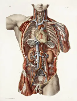
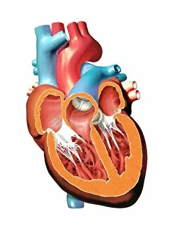
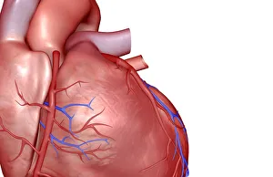
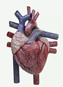
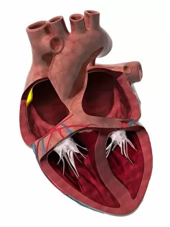
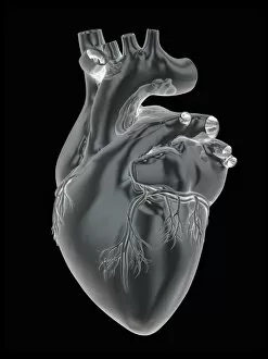
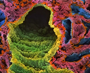
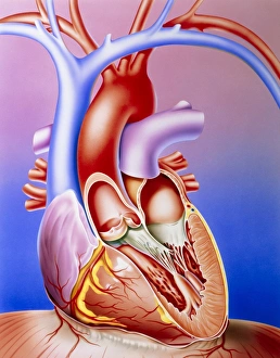
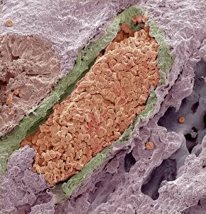
The pulmonary vein, a vital component of the cardiovascular system, is beautifully depicted in historical artwork showcasing the intricate anatomy of the human heart


All Professionally Made to Order for Quick Shipping
The pulmonary vein, a vital component of the cardiovascular system, is beautifully depicted in historical artwork showcasing the intricate anatomy of the human heart. These captivating illustrations not only highlight the structure and function of this crucial vessel but also provide insight into our understanding of the circulatory system. In these remarkable artworks, we can observe how the pulmonary vein connects directly to the left atrium of the heart. It serves as a conduit for oxygenated blood from the lungs back to this chamber, completing an essential loop in circulation between our heart and lungs. Furthermore, these artistic representations often showcase other significant elements such as the coronary vessels that nourish and supply oxygen-rich blood to different parts of our hearts. The intricate network formed by these vessels ensures proper functioning and health of this vital organ. One cannot help but marvel at detailed cross-section biomedical illustrations depicting alveoli – tiny air sacs within our lungs where gas exchange occurs. This connection between lung tissue and pulmonary veins further emphasizes their role in maintaining a healthy respiratory system. Artistic depictions also shed light on various lobes within our lungs, highlighting their interconnectedness with both arteries and veins. Such visualizations aid us in comprehending how blood flows through these complex structures while facilitating efficient gas exchange. Models representing human hearts offer another perspective on how all its chambers work together harmoniously with specialized nodes like sinus node (artwork C014 / 2030). These images allow us to appreciate not only the beauty but also complexity behind each heartbeat driven by electrical impulses originating from specific regions within our hearts. Exploring historical artwork portraying pulmonary veins alongside other components like coronary vessels or lung lobes provides valuable insights into both cardiac anatomy and overall circulatory dynamics. These captivating visuals serve as reminders of just how intricately designed our bodies are while fueling curiosity about ongoing advancements in cardiovascular research.