Fraud Blocker Skip to main content

Proteomics Collection (page 3)

Proteomics, the study of proteins and their functions within an organism, is a fascinating field that unravels the intricate workings of life

Background imageProteomics Collection: Human 80S ribosome F007 / 9898

Human 80S ribosome F007 / 9898
Ribosomal subunit. Computer model showing the structure of the RNA (ribonucleic acid) molecules in an 80S (large) ribosomal sub-unit. Ribosomes are composed of protein and RNA

Background imageProteomics Collection: Type I topoisomerase protein bound to DNA F007 / 9893

Type I topoisomerase protein bound to DNA F007 / 9893
Type I topoisomerase bound to DNA. Molecular model showing a type I topoisomerase molecule (blue) bound to a strand of DNA (deoxyribonucleic acid, yellow and red)

Background imageProteomics Collection: Ubiquitin activating enzyme protein E1 F007 / 9919

Ubiquitin activating enzyme protein E1 F007 / 9919
Molecular model of a ubiquitin-activating enzyme, also known as E1 enzymes. These catalyse the first step in the ubiquitination reaction, which targets a protein for degradation via a proteasome

Background imageProteomics Collection: Yeast enzyme, molecular model F007 / 9887

Yeast enzyme, molecular model F007 / 9887
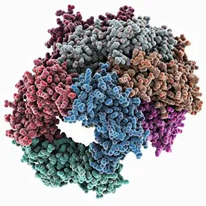
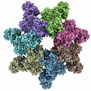
Yeast enzyme. Molecular model of an enzyme from bakers yeast (Saccharomyces cerevisiae). This is the 20S proteasome. A proteasome is a complex type of proteinase (protein-digesting enzyme)

Background imageProteomics Collection: DNA molecule, artwork F008 / 2034

DNA molecule, artwork F008 / 2034
DNA molecule, computer artwork

Background imageProteomics Collection: DNA molecule, artwork F008 / 2036

DNA molecule, artwork F008 / 2036
DNA molecule, computer artwork

Background imageProteomics Collection: DNA molecule, artwork F008 / 2040

DNA molecule, artwork F008 / 2040
DNA molecule, computer artwork

Background imageProteomics Collection: HIV-1 protease molecule

HIV-1 protease molecule
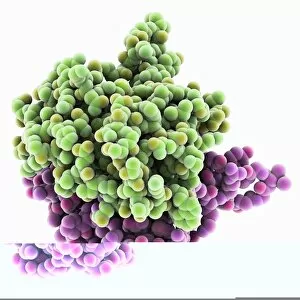
HIV-1 protease, molecular model. This enzyme, from HIV (human immunodeficiency virus), cleaves viral polyproteins into functional proteins that are essential for viral assembly and infectivity

Background imageProteomics Collection: HK97 bacteriophage capsid

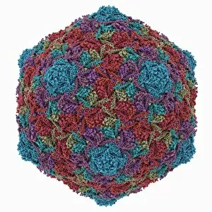
HK97 bacteriophage capsid, molecular model. Bacteriophages are viruses that infect bacteria, in this case enterobacteria such as E. coli (Escherichia coli), with the phage head shown here

Background imageProteomics Collection: Chikungunya virus capsid

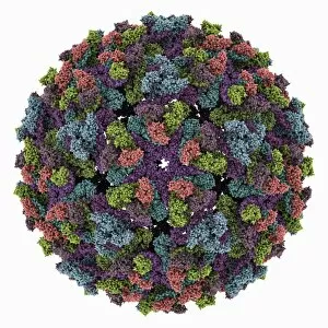
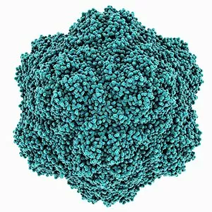
Chikungunya virus capsid, molecular model. This virus, transmitted by mosquitoes in tropical Africa and Asia, causes fever and joint pain in humans, similar to dengue fever

Background imageProteomics Collection: TATA box-binding protein complex C014 / 0867

TATA box-binding protein complex C014 / 0867
TATA box-binding protein complex. Molecular model showing a TATA box-binding protein (TBP, khaki) complexed with a strand of DNA (deoxyribonucleic acid)

Background imageProteomics Collection: HK97 bacteriophage procapsid

HK97 bacteriophage procapsid. Molecular model showing the structure of the prohead-I procapsid of the HK97 bacteriophage. Bacteriophages are viruses that infect bacteria

Background imageProteomics Collection: Turnip yellow mosaic virus capsid

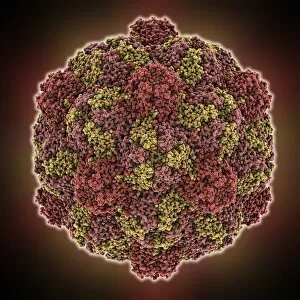
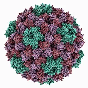
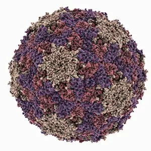
Turnip yellow mosaic virus capsid, molecular model. This virus infects a wide variety of plants, including crops such as turnips and cabbages, causing yellow patches on the leaves

Background imageProteomics Collection: Sindbis virus capsid, molecular model

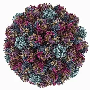
Sindbis virus capsid, molecular model. This virus, transmitted by mosquitoes, causes sindbis fever in humans. In viruses, the capsid is the protein shell that encloses the genetic material

Background imageProteomics Collection: Murine polyomavirus capsid

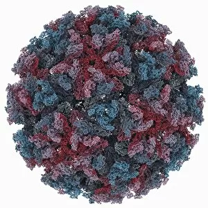
Murine polyomavirus capsid, molecular model. This virus, one of a range named for their potential to cause multiple tumours, infects mice

Background imageProteomics Collection: Human antitumour antibody molecule

Human antitumour antibody molecule
Human antitumour antibody. Molecular model showing the antitumour antibody BR96 complexed with part of the Lewis antigen. The Lewis antigen is expressed on the surface of human carcinoma cells

Background imageProteomics Collection: Iron containing protein, molecular model

Iron containing protein, molecular model
Iron containing protein. Molecular model showing the structure of a bacterial homolog of the animal iron containing protein ferritin

Background imageProteomics Collection: Brome mosaic virus capsid

Brome mosaic virus capsid, molecular model. This plant virus infects grasses, especially brome grasses, and also barley. It causes mosaic patches of discolouration

Background imageProteomics Collection: Cowpea chlorotic mottle virus capsid

Cowpea chlorotic mottle virus capsid, molecular model. This virus (CCMV) infects the cowpea plant (Vigna unguiculata), causing yellow spots of discolouration

Background imageProteomics Collection: Potassium ion channel protein structure

Potassium ion channel protein structure. Molecular model of a KcsA potassium ion (K+) channel from Streptomyces lividans bacteria

Background imageProteomics Collection: Potassium ion channel beta subunit

Potassium ion channel beta subunit. Molecular model showing the structure a beta subunit of a voltage-dependent potassium (K+) channel

Background imageProteomics Collection: Tyrosyl-tRNA synthetase molecule

Tyrosyl-tRNA synthetase molecule
Tyrosyl-tRNA synthetase protein molecule. Molecular model showing bacterial tyrosyl-tRNA synthetase complexed with tyrosyl tRNA (transfer ribonucleic acid)

Background imageProteomics Collection: KCNQ ion channel protein structure

KCNQ ion channel protein structure. Molecular model showing the protein structure of an ion channel domain. Ion channels are membrane-spanning proteins that form a pathway for the movement of

Background imageProteomics Collection: Proliferating cell nuclear antigen

Proliferating cell nuclear antigen molecule. Molecular model of human proliferating cell nuclear antigen (PCNA, blue, green and red), complexed with its loader protein (purple, orange)

Background imageProteomics Collection: Potassium ion channel cavity structure

Potassium ion channel cavity structure. Molecular model showing the structure of a cavity formed by potassium ion channel proteins

Background imageProteomics Collection: Tumour suppressor protein and DNA C017 / 3645

Tumour suppressor protein and DNA C017 / 3645
Tumour suppressor protein and DNA. Computer artwork showing a molecule of the tumour suppressor protein p53 (blue and pink) bound to a molecule of DNA (deoxyribonucleic acid, yellow and orange)

Background imageProteomics Collection: ATP synthase molecule C014 / 0880

ATP synthase molecule C014 / 0880
ATP synthase molecule. Molecular model showing the structure of ATP synthase (ATPase) subunit C. ATPase is an important enzyme that provides energy for cells through the synthesis of adenosine

Background imageProteomics Collection: Avian polyomavirus capsid

Avian polyomavirus capsid, molecular model. This virus, one of a range named for their potential to cause multiple tumours, infects birds. Discovered in budgerigars in 1981, it is often fatal

Background imageProteomics Collection: Anthrax protective antigen molecule C014 / 0886

Anthrax protective antigen molecule C014 / 0886
Anthrax protective antigen molecule. Computer model showing the structure of a molecule of protective antigen (PA) produced by anthrax (Bacillus anthracis) bacteria

Background imageProteomics Collection: HIV enzyme protein, molecular model C014 / 0876

HIV enzyme protein, molecular model C014 / 0876
HIV enzyme protein. Computer model showing the structure of the catalytic domain of a molecule of HIV-1 retroviral integrase (IN) from the human immunodeficiency virus (HIV)

Background imageProteomics Collection: Tryptophanyl-tRNA synthetase molecule

Tryptophanyl-tRNA synthetase molecule
Tryptophanyl-tRNA synthetase protein molecule. Molecular model showing human tryptophanyl-tRNA synthetase complexed with tryptophan tRNA (transfer ribonucleic acid)

Background imageProteomics Collection: Cytoplasmic polyhedrosis virus capsid

Cytoplasmic polyhedrosis virus capsid, molecular model. Part of the Cypovirus genus and invariably fatal, this insect virus is transmitted by contamination of leaves eaten (examples include silkworms)

Background imageProteomics Collection: Pho4 transcription factor bound to DNA C014 / 0861

Pho4 transcription factor bound to DNA C014 / 0861
Pho4 transcription factor bound to DNA. Molecular model showing phosphate system positive regulatory protein (Pho4) (blue and green) bound to a strand of DNA (deoxyribonucleic acid, red and purple)

Background imageProteomics Collection: DNA molecule, artwork F007 / 1996

DNA molecule, artwork F007 / 1996
DNA molecule, computer artwork

Background imageProteomics Collection: DNA molecule, artwork F007 / 1994

DNA molecule, artwork F007 / 1994
DNA molecule, computer artwork

Background imageProteomics Collection: DNA molecule, artwork F007 / 1995

DNA molecule, artwork F007 / 1995
DNA molecule, computer artwork

Background imageProteomics Collection: DNA molecule, artwork F007 / 1991

DNA molecule, artwork F007 / 1991
DNA molecule, computer artwork

Background imageProteomics Collection: DNA molecule, artwork F007 / 1992

DNA molecule, artwork F007 / 1992
DNA molecule, computer artwork

Background imageProteomics Collection: HIV enzyme protein, molecular model

HIV enzyme protein, molecular model
HIV enzyme protein. Computer model showing the structure of the catalytic domain of a molecule of HIV-1 retroviral integrase (IN) from the human immunodeficiency virus (HIV)

Background imageProteomics Collection: Anthrax protective antigen molecule C014 / 0865

Anthrax protective antigen molecule C014 / 0865
Anthrax protective antigen molecule. Computer model showing the structure of a molecule of protective antigen (PA) produced by anthrax (Bacillus anthracis) bacteria

Background imageProteomics Collection: TATA box-binding protein complex C017 / 7090

TATA box-binding protein complex C017 / 7090
TATA box-binding protein complex. Molecular model showing a TATA box-binding protein (TBP, green) complexed with a strand of DNA (deoxyribonucleic acid, spheres) and transcription factor IIB

Background imageProteomics Collection: Theilers encephalomyelitis virus capsid

Theilers encephalomyelitis virus capsid, molecular model. This virus, which causes brain and spinal cord inflammation in mice, is used in research

Background imageProteomics Collection: TATA box-binding protein complex C017 / 7085

TATA box-binding protein complex C017 / 7085
TATA box-binding protein complex. Molecular model showing a TATA box-binding protein (TBP, green) complexed with a strand of DNA (deoxyribonucleic acid, yellow) and transcription factor IIB

Background imageProteomics Collection: Adenine molecule, artwork C017 / 7199

Adenine molecule, artwork C017 / 7199
Adenine molecule. Computer artwork showing the structure of a molecule of the nucleobase adenine. Atoms are colour-coded spheres: carbon (green), nitrogen (blue), and oxygen (white)

Background imageProteomics Collection: Ricin A-chain, artwork C017 / 3654

Ricin A-chain, artwork C017 / 3654
Ricin A-chain. Computer artwork showing the enzymatically active A-chain from a molecule of the toxic protein ricin. Ricin comprises two entwined amino acid chains; A (seen here) and B (not shown)

Background imageProteomics Collection: Sirtuin enzyme and p53, artwork C017 / 3660

Sirtuin enzyme and p53, artwork C017 / 3660
Sirtuin enzyme and p53. Computer artwork of a sirtuin (Sir2) enzyme (blue) bound to a p53 peptide (pink). Sir2 enzymes form a unique class of NAD(+)

Background imageProteomics Collection: Tobacco necrosis virus capsid

Tobacco necrosis virus capsid, molecular model. This plant virus infects a wide rage of plants, including the tobacco plant for which it is named. The virus causes tissue death (necrosis)

Background imageProteomics Collection: Ricin molecule, artwork C017 / 3649

Ricin molecule, artwork C017 / 3649
Ricin molecule. Computer artwork showing the structure of a molecule of the toxic protein ricin. Ricin comprises two entwined amino acid chains; A (yellow) and B (blue)


All Professionally Made to Order for Quick Shipping

Proteomics, the study of proteins and their functions within an organism, is a fascinating field that unravels the intricate workings of life. From anaesthetics inhibiting ion channels to immunoglobulin G antibody molecules, proteomics delves into the molecular mechanisms that shape our existence. In the realm of brain research, scientists explore how proteins influence cognition and behavior. They investigate DNA nucleosomes' structure and function, unraveling their role in gene regulation. Antibodies take center stage as artwork showcases their diverse forms and crucial role in immune defense. Zinc fingers bound to a DNA strand highlight protein-DNA interactions critical for genetic processes. Meanwhile, manganese superoxide dismutase enzyme aids in protecting cells from oxidative stress. The SARS coronavirus protein becomes a subject of intense scrutiny as researchers strive to understand its pathogenicity. Cytochrome b5 molecule reveals insights into electron transfer reactions within cells while glutamine synthetase enzyme plays a vital role in nitrogen metabolism. Lastly, RNA-editing enzymes offer potential therapeutic targets for various diseases with their ability to modify genetic information at the RNA level. Through proteomics, we unlock nature's secrets one protein at a time - deciphering their structures, unraveling their functions, and ultimately enhancing our understanding of life itself.