Fraud Blocker Skip to main content

Proteins Collection (page 3)

Proteins: The Building Blocks of Life From the intricate network of nerve and glial cells to the mesmerizing patterns seen under a light micrograph

Background imageProteins Collection: Tumour suppressor protein and DNA C017 / 3647

Tumour suppressor protein and DNA C017 / 3647
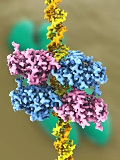
Tumour suppressor protein and DNA. Computer artwork showing a molecule of the tumour suppressor protein p53 (blue and pink) bound to a molecule of DNA (deoxyribonucleic acid, yellow and orange)
Background imageProteins Collection: TATA box-binding protein complex C017 / 7082

TATA box-binding protein complex C017 / 7082
TATA box-binding protein complex. Molecular model showing a TATA box-binding protein (TBP, green) complexed with a strand of DNA (deoxyribonucleic acid, yellow) and transcription factor IIB

Background imageProteins Collection: TATA box-binding protein complex C017 / 7088

TATA box-binding protein complex C017 / 7088
TATA box-binding protein complex. Molecular model showing a TATA box-binding protein (TBP, green) complexed with a strand of DNA (deoxyribonucleic acid, yellow) and transcription factor IIB

Background imageProteins Collection: GAL4p activator protein C017 / 7009

GAL4p activator protein C017 / 7009
Molecular structure of the Gal4p activator protein. It consists of two Gal4p, bound to a GAL upstream activator sequence (UAS)

Background imageProteins Collection: GAL4p activator protein C017 / 7008

GAL4p activator protein C017 / 7008
Molecular structure of the Gal4p activator protein. It consists of two Gal4p, bound to a GAL upstream activator sequence (UAS)

Background imageProteins Collection: TATA box-binding protein complex C017 / 7084

TATA box-binding protein complex C017 / 7084
TATA box-binding protein complex. Molecular model showing a TATA box-binding protein (TBP, green) complexed with a strand of DNA (deoxyribonucleic acid, yellow) and transcription factor IIB

Background imageProteins Collection: Sirtuin enzyme and p53, artwork C017 / 3659

Sirtuin enzyme and p53, artwork C017 / 3659
Sirtuin enzyme and p53. Computer artwork of a sirtuin (Sir2) enzyme (pink) bound to a p53 peptide (orange). Sir2 enzymes form a unique class of NAD(+)

Background imageProteins Collection: Adenine molecule, artwork C017 / 7200

Adenine molecule, artwork C017 / 7200
Adenine molecule. Computer artwork showing the structure of a molecule of the nucleobase adenine. Atoms are colour-coded spheres: carbon (green), nitrogen (blue), and oxygen (white)

Background imageProteins Collection: Tumour suppressor protein and DNA C017 / 3644

Tumour suppressor protein and DNA C017 / 3644
Tumour suppressor protein and DNA. Computer artwork showing a molecule of the tumour suppressor protein p53 (blue and pink) bound to a molecule of DNA (deoxyribonucleic acid, yellow and orange)

Background imageProteins Collection: Sirtuin enzyme and p53, artwork C017 / 3658

Sirtuin enzyme and p53, artwork C017 / 3658
Sirtuin enzyme and p53. Computer artwork of a sirtuin (Sir2) enzyme (pink) bound to a p53 peptide (orange). Sir2 enzymes form a unique class of NAD(+)

Background imageProteins Collection: SIRT3 molecule, artwork C017 / 3657

SIRT3 molecule, artwork C017 / 3657
SIRT3 molecule. Computer artwork showing the structure of a molecule of NAD-dependent deacetylase sirtuin-3, mitochondrial (SIRT3)

Background imageProteins Collection: Tumour suppressor protein and DNA C017 / 3646

Tumour suppressor protein and DNA C017 / 3646
Tumour suppressor protein and DNA. Computer artwork showing a molecule of the tumour suppressor protein p53 (blue and pink) bound to a molecule of DNA (deoxyribonucleic acid, yellow and orange)

Background imageProteins Collection: Thymine molecule, artwork C017 / 7366

Thymine molecule, artwork C017 / 7366
Thymine molecule. Computer artwork showing the structure of a molecule of the nucleobase thymine. Atoms are colour-coded spheres: carbon (green), nitrogen (blue), oxygen (red), and hydrogen (white)

Background imageProteins Collection: Thymine molecule, artwork C017 / 7365

Thymine molecule, artwork C017 / 7365
Thymine molecule. Computer artwork showing the structure of a molecule of the nucleobase thymine. Atoms are colour-coded spheres: carbon (green), nitrogen (blue), oxygen (red), and hydrogen (white)

Background imageProteins Collection: Heat shock factor protein F007 / 9885

Heat shock factor protein F007 / 9885
Molecular model of a Heat Shock Protein (HSP).HSPs are a group of proteins whose levels increase when cells are exposed to raised temperatures or other stress

Background imageProteins Collection: Heat shock factor 70 protein F007 / 9895

Heat shock factor 70 protein F007 / 9895
Molecular model of the Heat Shock Protein 70 (HSP).HSPs are a group of proteins whose levels increase when cells are exposed to raised temperatures or other stress

Background imageProteins Collection: Haemagglutinin viral surface protein F007 / 9932

Haemagglutinin viral surface protein F007 / 9932
Haemagglutinin viral surface protein. Molecular model of haemagglutinin, a surface protein from the influenza virus, complexed with a neutralising antibody

Background imageProteins Collection: Haemagglutinin viral surface protein F007 / 9931

Haemagglutinin viral surface protein F007 / 9931
Haemagglutinin viral surface protein. Molecular model of haemagglutinin, a surface protein from the influenza virus, complexed with a neutralising antibody

Background imageProteins Collection: DNA nucleosome, molecular model F007 / 9883

DNA nucleosome, molecular model F007 / 9883
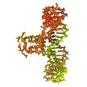
DNA nucleosome. Molecular model of a nucleosome, the fundamental repeating unit used to package DNA (deoxyribonucleic acid) inside cell nuclei

Background imageProteins Collection: DNA nucleosome, molecular model F007 / 9888

DNA nucleosome, molecular model F007 / 9888
DNA nucleosome. Molecular model of a nucleosome, the fundamental repeating unit used to package DNA (deoxyribonucleic acid) inside cell nuclei

Background imageProteins Collection: Heat shock factor 70 protein F007 / 9903

Heat shock factor 70 protein F007 / 9903
Molecular model of the Heat Shock Protein 70 (HSP).HSPs are a group of proteins whose levels increase when cells are exposed to raised temperatures or other stress

Background imageProteins Collection: Human serum albumin molecule F007 / 9904

Human serum albumin molecule F007 / 9904
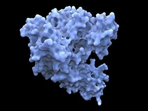
Human serum albumin, molecular model. Albumin is the most abundant protein in human blood plasma. One of albumins functions is to transport fatty acids to the liver

Background imageProteins Collection: Human 80S ribosome F007 / 9902

Human 80S ribosome F007 / 9902
Ribosomal subunit. Computer model showing the structure of the RNA (ribonucleic acid) molecules in an 80S (large) ribosomal sub-unit. Ribosomes are composed of protein and RNA

Background imageProteins Collection: Heat shock factor protein F007 / 9892

Heat shock factor protein F007 / 9892
Molecular model of a Heat Shock Protein (HSP).HSPs are a group of proteins whose levels increase when cells are exposed to raised temperatures or other stress

Background imageProteins Collection: Human serum albumin molecule F007 / 9905

Human serum albumin molecule F007 / 9905
Human serum albumin, molecular model. Albumin is the most abundant protein in human blood plasma. One of albumins functions is to transport fatty acids to the liver

Background imageProteins Collection: Human 80S ribosome F007 / 9898

Human 80S ribosome F007 / 9898
Ribosomal subunit. Computer model showing the structure of the RNA (ribonucleic acid) molecules in an 80S (large) ribosomal sub-unit. Ribosomes are composed of protein and RNA

Background imageProteins Collection: Type I topoisomerase protein bound to DNA F007 / 9893

Type I topoisomerase protein bound to DNA F007 / 9893
Type I topoisomerase bound to DNA. Molecular model showing a type I topoisomerase molecule (blue) bound to a strand of DNA (deoxyribonucleic acid, yellow and red)

Background imageProteins Collection: Balanced diet, conceptual image F005 / 0358

Balanced diet, conceptual image F005 / 0358
Balanced diet, conceptual image

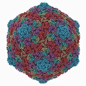
Background imageProteins Collection: HK97 bacteriophage capsid

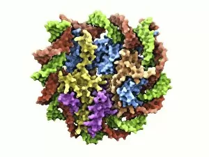
HK97 bacteriophage capsid, molecular model. Bacteriophages are viruses that infect bacteria, in this case enterobacteria such as E. coli (Escherichia coli), with the phage head shown here

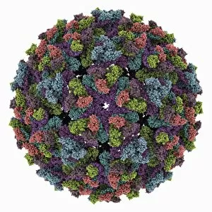
Background imageProteins Collection: Chikungunya virus capsid

Chikungunya virus capsid, molecular model. This virus, transmitted by mosquitoes in tropical Africa and Asia, causes fever and joint pain in humans, similar to dengue fever

Background imageProteins Collection: TATA box-binding protein complex C014 / 0867

TATA box-binding protein complex C014 / 0867
TATA box-binding protein complex. Molecular model showing a TATA box-binding protein (TBP, khaki) complexed with a strand of DNA (deoxyribonucleic acid)

Background imageProteins Collection: HK97 bacteriophage procapsid

HK97 bacteriophage procapsid. Molecular model showing the structure of the prohead-I procapsid of the HK97 bacteriophage. Bacteriophages are viruses that infect bacteria

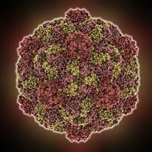
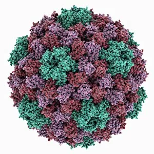
Background imageProteins Collection: Turnip yellow mosaic virus capsid

Turnip yellow mosaic virus capsid, molecular model. This virus infects a wide variety of plants, including crops such as turnips and cabbages, causing yellow patches on the leaves

Background imageProteins Collection: Sindbis virus capsid, molecular model

Sindbis virus capsid, molecular model. This virus, transmitted by mosquitoes, causes sindbis fever in humans. In viruses, the capsid is the protein shell that encloses the genetic material

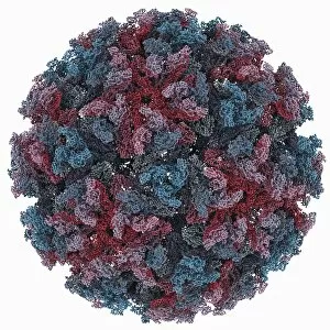
Background imageProteins Collection: Murine polyomavirus capsid

Murine polyomavirus capsid, molecular model. This virus, one of a range named for their potential to cause multiple tumours, infects mice

Background imageProteins Collection: Iron containing protein, molecular model

Iron containing protein, molecular model
Iron containing protein. Molecular model showing the structure of a bacterial homolog of the animal iron containing protein ferritin

Background imageProteins Collection: Brome mosaic virus capsid

Brome mosaic virus capsid, molecular model. This plant virus infects grasses, especially brome grasses, and also barley. It causes mosaic patches of discolouration

Background imageProteins Collection: Cowpea chlorotic mottle virus capsid

Cowpea chlorotic mottle virus capsid, molecular model. This virus (CCMV) infects the cowpea plant (Vigna unguiculata), causing yellow spots of discolouration

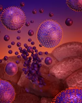
Background imageProteins Collection: Flu virus infection, conceptual image

Flu virus infection, conceptual image. Computer artwork showing influenza virus particles (round) in the respiratory tract

Background imageProteins Collection: Potassium ion channel protein structure

Potassium ion channel protein structure. Molecular model of a KcsA potassium ion (K+) channel from Streptomyces lividans bacteria

Background imageProteins Collection: Potassium ion channel beta subunit

Potassium ion channel beta subunit. Molecular model showing the structure a beta subunit of a voltage-dependent potassium (K+) channel

Background imageProteins Collection: Stem cell-derived retinal cells

Stem cell-derived retinal cells. Fluorescence light micrograph of retinal pigment epithelium (RPE) cells that have been derived from human embryonic stem cells (HESC)

Background imageProteins Collection: Tyrosyl-tRNA synthetase molecule

Tyrosyl-tRNA synthetase molecule
Tyrosyl-tRNA synthetase protein molecule. Molecular model showing bacterial tyrosyl-tRNA synthetase complexed with tyrosyl tRNA (transfer ribonucleic acid)

Background imageProteins Collection: KCNQ ion channel protein structure

KCNQ ion channel protein structure. Molecular model showing the protein structure of an ion channel domain. Ion channels are membrane-spanning proteins that form a pathway for the movement of

Background imageProteins Collection: Proliferating cell nuclear antigen

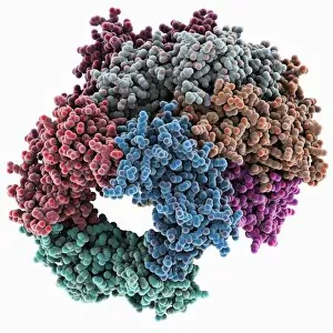
Proliferating cell nuclear antigen molecule. Molecular model of human proliferating cell nuclear antigen (PCNA, blue, green and red), complexed with its loader protein (purple, orange)

Background imageProteins Collection: Potassium ion channel cavity structure

Potassium ion channel cavity structure. Molecular model showing the structure of a cavity formed by potassium ion channel proteins

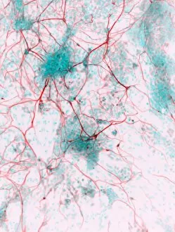
Background imageProteins Collection: Stem cell-derived neurons, micrograph

Stem cell-derived neurons, micrograph
Stem cell-derived neurons. Light micrograph of human nerve cells (neurons) that have been derived from induced pluripotent stem cells (IPS). Tuj1 proteins are cyan, and cell nuclei are red

Background imageProteins Collection: Tumour suppressor protein and DNA C017 / 3645

Tumour suppressor protein and DNA C017 / 3645
Tumour suppressor protein and DNA. Computer artwork showing a molecule of the tumour suppressor protein p53 (blue and pink) bound to a molecule of DNA (deoxyribonucleic acid, yellow and orange)


All Professionally Made to Order for Quick Shipping

Proteins: The Building Blocks of Life From the intricate network of nerve and glial cells to the mesmerizing patterns seen under a light micrograph, proteins play an essential role in every aspect of our existence, and are like the conductors of our body's symphony, orchestrating vital processes that keep us alive and functioning. Take, for example, an anaesthetic inhibiting an ion channel C015 / 6718. Proteins act as gatekeepers, controlling what enters or exits our cells. In this case, they regulate the flow of ions necessary for transmitting nerve signals and maintaining proper cell function. But proteins don't just govern our internal workings; they also interact with external threats such as the avian flu virus. These microscopic invaders hijack host cells using their own protein machinery to replicate themselves. Understanding these interactions is crucial in developing effective treatments against viral infections. While some proteins protect us from harm, others contribute to overall well-being through a balanced diet. Our bodies require various types found in different foods to ensure optimal health and nutrition. The secondary structure is truly a work of art—a complex folding pattern that determines their shape and function. Artists have captured this beauty through stunning artwork showcasing these intricate molecular structures. One such structure is the nucleosome molecule—an elegant arrangement where DNA wraps around protein spools called histones—forming compact units within chromosomes. This organization allows efficient storage and retrieval of genetic information during cell division or gene expression. Antibodies are another remarkable class depicted in captivating artwork. These specialized molecules recognize foreign substances like bacteria or viruses and neutralize them by binding tightly to specific targets on their surface—an extraordinary defense mechanism employed by our immune system. Speaking of bacteria, their ribosomes serve as factories producing new proteins based on instructions encoded in DNA—the blueprint for life itself. Understanding bacterial ribosomes has led to groundbreaking discoveries in antibiotic development, combating infectious diseases that threaten human health.