
Prokaryote Collection
Prokaryotes: The Marvels of Microscopic Life Cell types, artwork, and a world unseen come together to unravel the fascinating realm of prokaryotes


All Professionally Made to Order for Quick Shipping
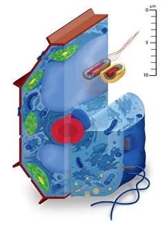
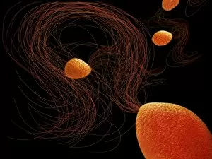
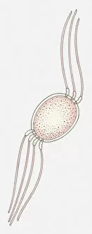
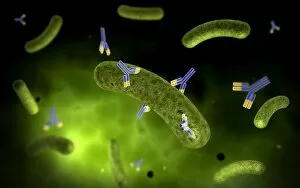
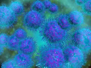
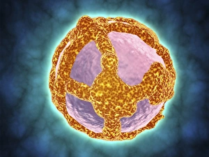
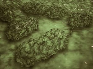
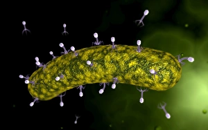
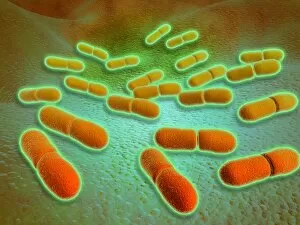
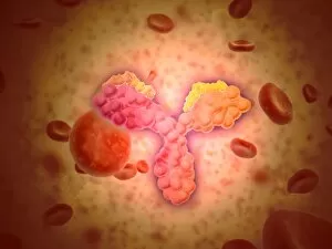
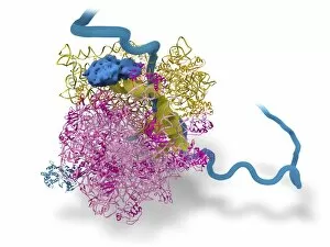
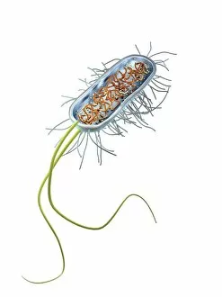
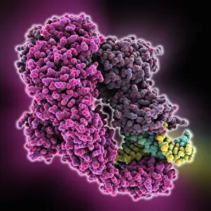
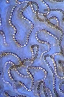
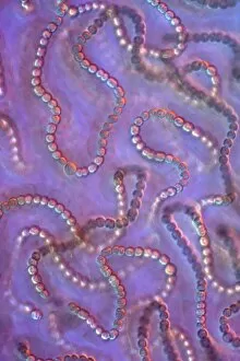
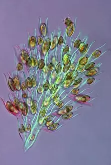
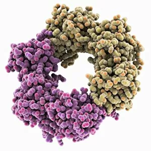
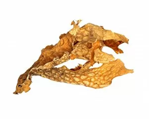
Prokaryotes: The Marvels of Microscopic Life Cell types, artwork, and a world unseen come together to unravel the fascinating realm of prokaryotes. From Pyrococcus furiosus archaea artwork to Bacillus bacterial genus illustrations, these captivating visuals shed light on the diversity within this ancient group of organisms. Peering through a microscope, we witness the intricate beauty cells. A stunning illustration showcases their simplicity yet complexity—a reminder that life can thrive in even the tiniest packages. Microscopic views reveal streptococcus bacteria forming chains like delicate pearls while an antibody attaches itself to a bacterium, executing its lethal mission. Diplococcus bacterium appears as pairs under scrutiny, showcasing their unique structure and form. Intriguingly named Listeria monocytogenes captures our attention with its microscopic view—an organism that challenges our understanding of survival and adaptation. Meanwhile, electron micrographs unveil Prochlorococcus—nature's tiny photosynthetic powerhouse—giving us insight into how it harnesses sunlight for energy production. A group portrait reveals Borrelia burgdorferi bacteria huddled together—a sight both mesmerizing and unsettling as they cause Lyme disease in humans. These glimpses into their hidden world remind us of nature's intricacies at play. Zooming out from individual species to encompass all bacteria brings forth an awe-inspiring perspective—the sheer multitude and variety that make up this vast microbial kingdom become apparent. Bacterial pneumonia is captured in microscopic detail—a stark reminder of both their potential harm and vital role in shaping ecosystems, and are not just subjects for scientific study; they are living masterpieces waiting to be explored further. Through artful depictions or microscopic revelations, we uncover the wonders concealed within these minuscule beings—the architects of life on Earth since time immemorial.