
Plasma Membrane Collection
The plasma membrane, also known as the cell membrane, is a vital component of various cells in living organisms


All Professionally Made to Order for Quick Shipping
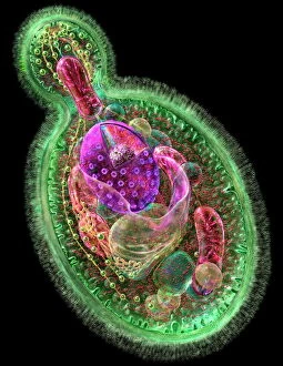
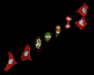
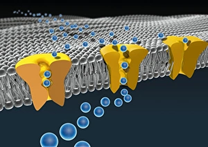
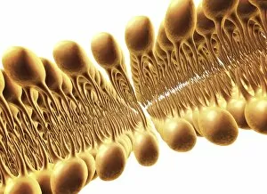
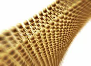
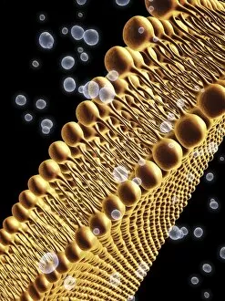
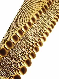
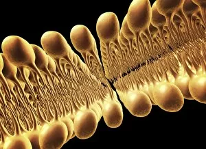
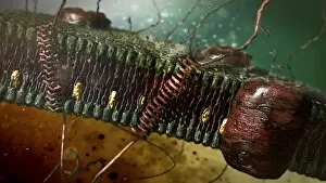
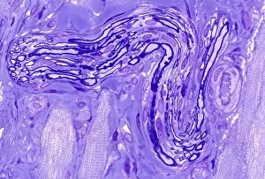
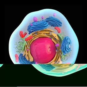
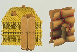
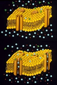
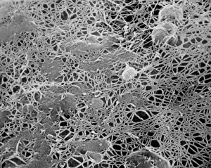
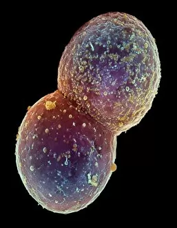
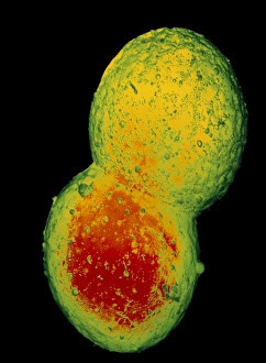
The plasma membrane, also known as the cell membrane, is a vital component of various cells in living organisms. In budding yeast cells, this thin barrier plays a crucial role in maintaining cellular integrity and regulating the exchange of molecules with its surroundings. During mitosis, when cells divide to form new ones, the plasma membrane undergoes intricate changes that ensure proper distribution of genetic material. This process can be visualized under a light micrograph, revealing the dynamic nature of this essential structure. Artwork C013 / 7467 beautifully captures the complexity of the cell membrane's lipid bilayer composition. Composed of phospholipids and proteins, this double-layered arrangement acts as a selective barrier that controls what enters or exits the cell. Artwork F007 / 1477 showcases another aspect of the plasma membrane – ion channels. These specialized protein structures allow ions to pass through selectively, facilitating crucial cellular processes such as nerve impulse transmission and muscle contraction. In an intricately depicted muscle fiber structure artwork, we witness how tightly packed plasma membranes contribute to muscular function by enabling coordinated contractions for movement and strength. A light micrograph showcasing bladder epithelium reveals yet another example where they are critical for maintaining tissue integrity and preventing harmful substances from entering our bodies. A detailed model of an animal cell highlights numerous organelles within its cytoplasm but prominently features a thin plasma membrane adorned with microvilli projections at its top surface. These finger-like extensions increase surface area for enhanced nutrient absorption or sensory reception in specialized cells like those lining our intestines. Additional artwork (F007 / 1479-1480) emphasizes different aspects of lipid bilayers present in the cell membrane. The unique properties conferred by these lipids enable compartmentalization within cells while providing structural support and flexibility necessary for various cellular functions. Lastly, artwork F007 / 1478 showcases how closely packed lipids create tight junctions between adjacent cells, ensuring impermeability and maintaining tissue integrity.