
Planche Anatomique Collection
A planche anatomique is a detailed and informative illustration of the human body, showcasing the intricate systems and structures that make up our anatomy


All Professionally Made to Order for Quick Shipping
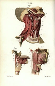
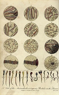
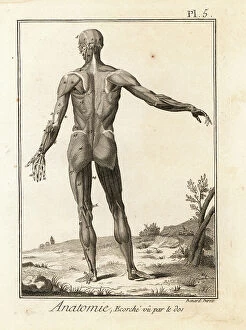
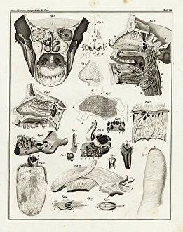
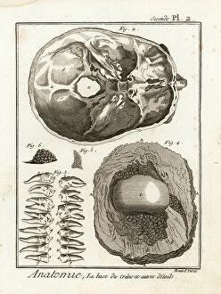
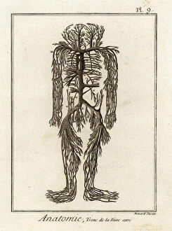
A planche anatomique is a detailed and informative illustration of the human body, showcasing the intricate systems and structures that make up our anatomy. These visual aids are commonly used in medical settings to educate students, healthcare professionals, and patients about various aspects of the body's physiology. From the skeletal system to the muscular system, a planche anatomique provides a comprehensive look at how our bodies function and interact with one another. By studying these diagrams, individuals can gain a deeper understanding of their own bodies and how different parts work together to maintain health and well-being. Whether you're learning about bones, organs, or nerves, a planche anatomique offers a clear and concise representation of each component. With vibrant colors and detailed labels, these illustrations make it easy to identify different parts of the body and understand their functions.