
Phalanx Collection (page 9)
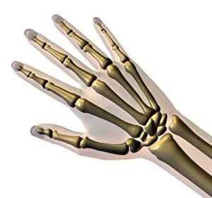
"Unveiling the Intricacies of the Phalanx: From Normal Foot to Inner Ankle Ligaments" In this captivating journey through human anatomy


All Professionally Made to Order for Quick Shipping
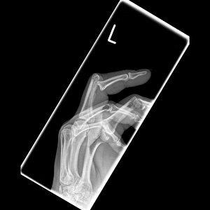
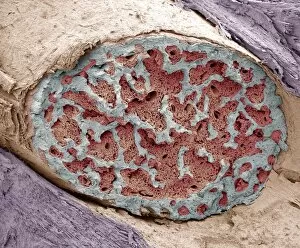
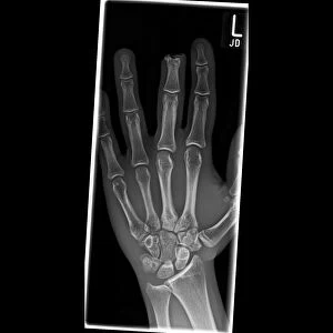
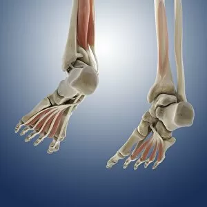
"Unveiling the Intricacies of the Phalanx: From Normal Foot to Inner Ankle Ligaments" In this captivating journey through human anatomy, we explore the fascinating world of the phalanx. Starting with a normal foot X-ray, we delve into the intricate layers that make up this vital part of our body. The human foot anatomy reveals a mesmerizing tapestry of skin, veins, arteries, muscles, and bones working in harmony. Moving on to outer ankle ligaments depicted in artwork C013/4452, we witness their strength and resilience as they support our every step. Similarly, inner ankle ligaments portrayed in artwork C013/4451 showcase their crucial role in maintaining stability and balance. Venturing beyond mere physicality, palmistry introduces us to a planetary and zodiacal diagram that adds an element of mystique to our exploration. Meanwhile, Francisco Franco's "Diario de una Bandera" cover reminds us of historical significance tied to symbols like flags. As we dive deeper into history's annals, iconic battles come alive before our eyes. From "The Battle of Waterloo, " immortalized on canvas by skilled artists capturing its intensity; to Pausanias' grandeur illustrated in Hutchinsons History; or even comparing hand bones across diverse mammals - each depiction unveils unique aspects related to warfare or evolution. Finally reaching ancient times when military formations reigned supreme comes "The Phalanx attacking the Centre on the Hydaspes. " This lithograph transports us back to those fierce encounters where unity and coordination were paramount for success. And who can forget "The Battle of Marathon, " another pivotal moment etched forever within Hutchinsons History? Concluding with a colored engraving showcasing "The Macedonians, " we realize how these warriors left an indelible mark on history through their strategic use of phalanxes.