
Phagocytosing Collection
"Unleashing the Power of Phagocytosis: A Fascinating Journey into Cellular Defense" In a microscopic battlefield


All Professionally Made to Order for Quick Shipping
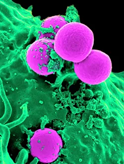
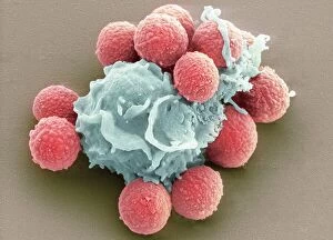
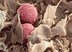
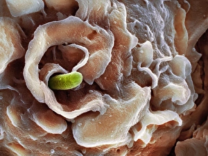
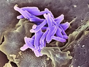
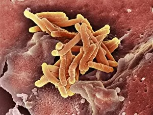
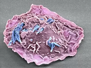
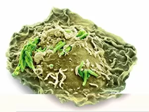
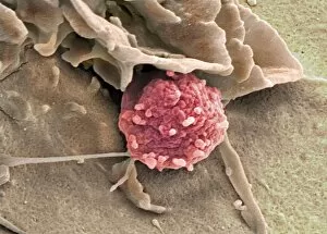
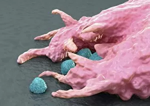
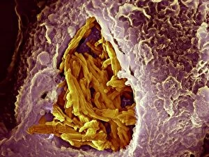
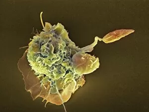
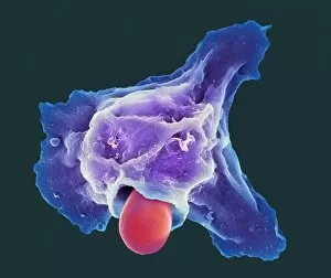
"Unleashing the Power of Phagocytosis: A Fascinating Journey into Cellular Defense" In a microscopic battlefield, neutrophils and macrophages emerge as mighty warriors against harmful invaders. Witness the extraordinary process of phagocytosis, where these immune cells engulf and devour pathogens to protect our bodies. Captured under the lens of a scanning electron microscope (SEM), we delve into this captivating world. Neutrophils are seen in action, fearlessly phagocytosing MRSA bacteria, their elongated pseudopods wrapping around the pathogen like a predator capturing its prey (Neutrophil engulfing MRSA, SEM C018 / 8596). But it doesn't stop there; fungi spores fall victim to this cellular defense mechanism too. The mesmerizing images reveal macrophages gracefully engulfing fungal spores with their intricate membrane extensions (Phagocytosis of fungal spores, SEM). This dance between host and invader showcases nature's remarkable ability to maintain equilibrium. The battle intensifies when tuberculosis bacteria come face-to-face with macrophages. In an awe-inspiring display of strength and resilience, these immune cells envelop TB bacteria within their cytoplasmic grasp (Macrophage engulfing TB bacteria, SEM). These images serve as a testament to the tireless efforts our body undertakes in combating infectious diseases. Shigella bacterium finds itself entangled in another gripping encounter with macrophages. The striking SEM captures moments frozen in time as these brave defenders capture Shigella within their clutches (Shigella bacterium and macrophage, SEM C016 / 8922-8924), and is through such battles that our immune system ensures our well-being remains intact. Even beyond bacterial foes lie ciliate protozoans ingesting algae—a reminder that phagocytic prowess extends across various organisms (Ciliate protozoan ingesting algae).