
Petri Dish Collection
In the world of microbiology, the a canvas for scientific exploration. It serves as a platform to cultivate and study various organisms, from bacteria to fungi


All Professionally Made to Order for Quick Shipping
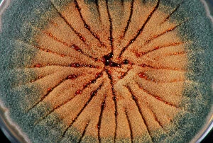
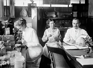
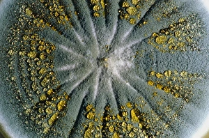
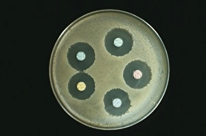
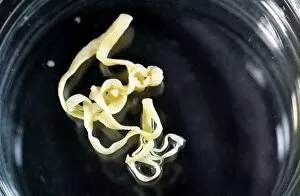
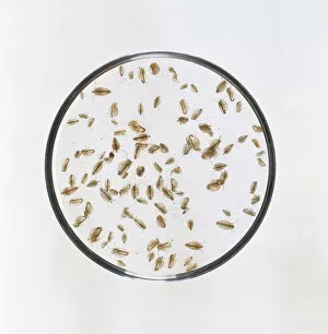
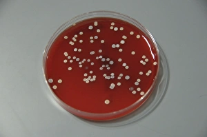
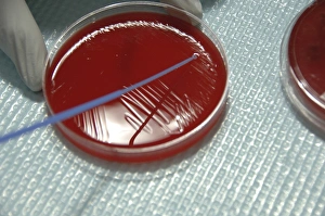
In the world of microbiology, the a canvas for scientific exploration. It serves as a platform to cultivate and study various organisms, from bacteria to fungi. One captivating example is the culture of Aspergillus nidulans fungus, which thrives within these glass plates. When peering into a petri dish under a microscope, one can witness mesmerizing patterns formed by Paenibacillus bacteria. These intricate structures are adaptive responses to laboratory-imposed stresses that mimic their natural environments. The C type exhibits chiral formations, while the T type showcases tip-splitting morphotypes. The significance of petri dishes extends beyond bacterial cultures; they also play an essential role in plant biotechnology research. Scientists utilize these vessels to propagate and manipulate plants for various purposes such as genetic modification or disease resistance studies. Looking back at history, we find iconic images associated with this scientific tool. A colorful lithograph from 1948 depicts a chemistry set and microscope - symbols of discovery and experimentation. Another black-and-white photograph captures Alexander Fleming himself around 1945, whose groundbreaking work on antibiotics revolutionized medicine. Even before modern times, pioneers like Oswald Theodore Avery paved the way for our understanding of microbial life through their tireless efforts in bacteriology and molecular biology. And let's not forget historical depictions showcasing antibiotic action against harmful bacteria during the nineteenth century. From Salmonella cultures to diverse bacterial colonies thriving on agar surfaces - petri dishes have become indispensable tools in unraveling nature's microscopic wonders.