
Pedicle Collection
"Exploring the Intricacies of Pedicle: From Kidney Glomerulus to Brachiopods" Delving into the microscopic world


All Professionally Made to Order for Quick Shipping
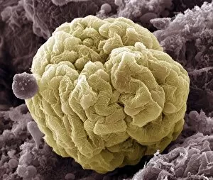
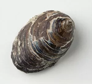
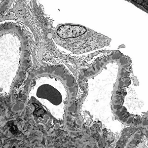
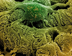
"Exploring the Intricacies of Pedicle: From Kidney Glomerulus to Brachiopods" Delving into the microscopic world, we observe a kidney glomerulus through the lens of a scanning electron microscope (SEM), revealing its intricate structure. Pancratium Speciosum, captured in Plate 47 from A Selection of Hexandrian Plants, showcases the delicate beauty of nature's pedicles. An exquisite illustration from 1831-1834 presents us with another glimpse into Pancratium Speciosum's pedicle, highlighting its unique characteristics. Shifting our focus to human anatomy, we explore the vertebra and uncover how it relies on pedicles for stability and support. Actinoconchus paradoxus McCoy demonstrates its attachment to hard substrates using a short pedicle; its brachial valve mirroring the surface it clings onto. Derbyia grandis (Waagen) reveals an intriguing free-living brachiopod with a remarkable pedicle valve that aids in locomotion and survival. Discinisca lugubris (Conrad) exhibits a conical pedicle valve within its shell, showcasing adaptations that enable this brachiopod species to thrive in various environments. Returning to renal wonders, we venture deeper into kidney glomeruli using transmission electron microscopy (TEM), unraveling their complex network responsible for filtration and waste removal. Nature never ceases to amaze as we zoom in on butterfly wing scale details through SEM imaging techniques - even these tiny structures possess fascinating pedicular arrangements. Our journey concludes by revisiting kidney glomeruli once more through SEM imagery; each capture emphasizing their vital role in maintaining proper kidney function and overall health.