Fraud Blocker Skip to main content

Pathogenic Collection

Pathogenic microorganisms are microscopic entities that pose a significant threat to human health

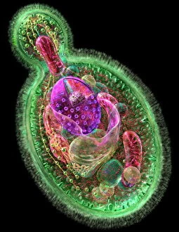
Background imagePathogenic Collection: Budding yeast cell

Budding yeast cell. Computer artwork of asection through a yeast ( Candida albicans )cell that is reproducing asexually. A daughtercell (top left) is budding from the parent cell

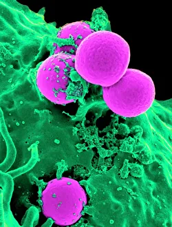
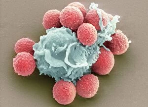
Background imagePathogenic Collection: Neutrophil engulfing MRSA, SEM C018 / 8596

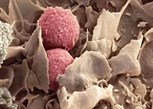
Neutrophil engulfing MRSA, SEM C018 / 8596
Neutrophil engulfing MRSA. Coloured scanning electron micrograph (SEM) of a neutrophil white blood cell (green) engulfing methicillin-resistant Staphylococcus aureus bacteria (MRSA, pink)

Background imagePathogenic Collection: E. coli bacteria, SEM

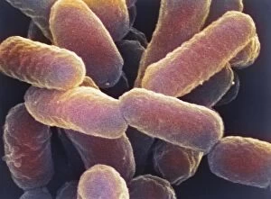
E. coli bacteria, SEM
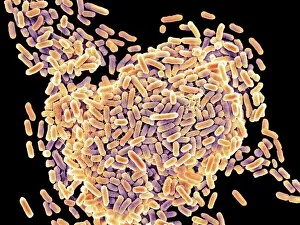
E. coli bacteria. Coloured scanning electron micrograph (SEM) of Escherichia coli bacteria (purple) taken from the small intestine of a child. E

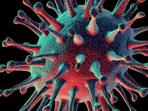
Background imagePathogenic Collection: Avian flu virus

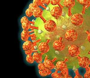
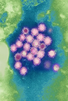
Avian flu virus, computer artwork. A virus is a tiny pathogenic particle comprising genetic material enclosed in a protein coat. The coat contains surface proteins (spikes)

Background imagePathogenic Collection: Salmonella bacteria, SEM

Salmonella bacteria, SEM
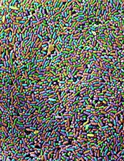
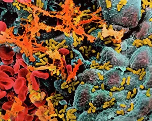
Salmonella bacteria, coloured scanning electron micrograph (SEM). These Gram-negative rod-shaped bacteria can cause food poisoning when eaten in contaminated food

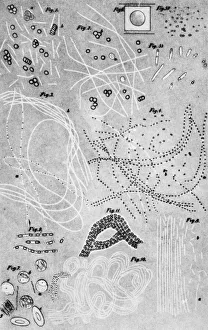
Background imagePathogenic Collection: Anthrax cultures, historical diagram

Anthrax cultures, historical diagram
Anthrax cultures. 1876 diagram of Bacillus anthracis (Anthrax) bacteria cultured by Robert Koch. Along with Louis Pasteur, Koch is considered the founder of modern medical bacteriology

Background imagePathogenic Collection: Salmonella bacteria, SEM

Salmonella bacteria, SEM
Salmonella bacteria, coloured scanning electron micrograph (SEM). Each rod-shaped structure is an individual bacterium. These bacteria occur mainly in human and animal intestines

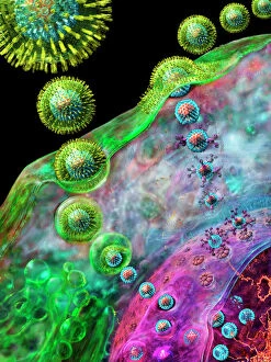
Background imagePathogenic Collection: Herpes virus replicating

Herpes virus replicating, computer artwork.Viruses are only able to replicate in a host cell.The glycoprotein spikes in the virus envelope(green)

Background imagePathogenic Collection: HIV particle

HIV particle, computer artwork. HIV (human immunodeficiency virus) causes AIDS (acquired immune deficiency syndrome). The virus consists of an RNA genome

Background imagePathogenic Collection: Avian flu virus

Avian flu virus, computer artwork. A virus is a tiny pathogenic particle comprising genetic material enclosed in a protein coat. The coat contains surface proteins (spikes)

Background imagePathogenic Collection: Norovirus particles, TEM

Norovirus particles, TEM
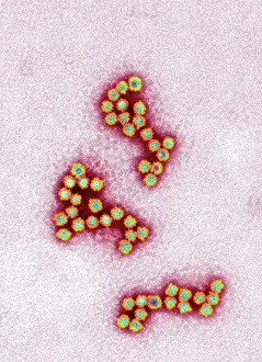
Norovirus particles. Coloured transmission electron micrograph (TEM) of norovirus particles. Norovirus is a genus of RNA (ribonucleic acid)

Background imagePathogenic Collection: Tuberculosis bacteria

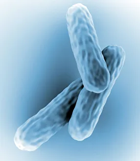
Tuberculosis bacteria. Computer artwork of Mycobacterium tuberculosis bacteria. These Gram- positive rod-shaped bacteria cause the disease tuberculosis

Background imagePathogenic Collection: Bacteria infecting a macrophage, SEM

Bacteria infecting a macrophage, SEM
Bacteria infecting a macrophage. Coloured scanning electron micrograph (SEM) of Mycobacterium tuberculosis bacteria (purple) infecting a macrophage white blood cell

Background imagePathogenic Collection: E. coli bacteria

E. coli bacteria
False-colour transmission electron micrograph of the bacterium Escherichia coli, a normal inhabitant of the human intestine

Background imagePathogenic Collection: Pseudomonas aeruginosa bacteria, SEM

Pseudomonas aeruginosa bacteria, SEM
Pseudomonas aeruginosa bacteria, coloured scanning electron micrograph (SEM). These Gram-negative rod-shaped bacteria are found in soil and water, and as normal flora in the human intestine

Background imagePathogenic Collection: E. coli bacterium, TEM

E. coli bacterium, TEM
E. coli bacterium, coloured transmission electron micrograph (TEM). E. coli are Gram-negative rod-shaped bacteria that are part of the normal flora of the human gut

Background imagePathogenic Collection: Hepatitis B viruses

Hepatitis B viruses
Hepatitis B virus. Coloured transmission electron micrograph (TEM) of hepatitis B viruses (circles), a cause of liver inflammation

Background imagePathogenic Collection: Helicobacter pylori bacteria

Helicobacter pylori bacteria
False-colour transmission electron micrograph of the bacterium Helicobacter pylori (formerly called Campylobacter pyloridis)

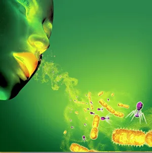
Background imagePathogenic Collection: Infections spread by sneezing, artwork C013 / 5949

Infections spread by sneezing, artwork C013 / 5949
Infections spread by sneezing. Computer artwork showing virus particles (virions, blue) and bacteria (rod-shaped) in the spray produced by someone when they sneeze

Background imagePathogenic Collection: Salmonella bacteria, artwork C013 / 8818

Salmonella bacteria, artwork C013 / 8818
Salmonella bacteria, computer artwork. Salmonella sp. bacteria are gram-negative rod-shaped bacteria that have flagella (hair-like structures) that they use for locomotion

Background imagePathogenic Collection: Phagocytosis of fungal spores, SEM

Phagocytosis of fungal spores, SEM
Phagocytosis of a fungus spore. Coloured scanning electron micrograph (SEM) of a neutrophil (blue) phagocytosing (engulfing and destroying) spores from the fungus Aspergillus fumigatus (red)

Background imagePathogenic Collection: Hepatitis B viruses

Hepatitis B viruses

Background imagePathogenic Collection: Influenza virus, computer artwork

Influenza virus, computer artwork
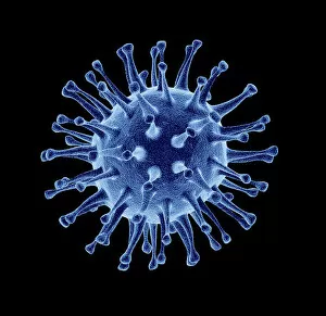
Influenza virus. Computer artwork of an influenza (flu) virus. The virus consists of a core of RNA (ribonucleic acid) genetic material surrounded by a protein coat

Background imagePathogenic Collection: Influenza virus particles, TEM

Influenza virus particles, TEM
Influenza virus particles, coloured transmission electron micrograph (TEM). This virus is the cause of the respiratory disease influenza (flu) in humans and other animals

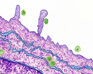
Background imagePathogenic Collection: Hepatitis C viruses, TEM

Hepatitis C viruses, TEM
Hepatitis C viruses. Coloured transmission electron micrograph (TEM) of hepatitis C virus particles (green) infecting cultured liver cells (purple)

Background imagePathogenic Collection: E. coli bacterium, TEM

E. coli bacterium, TEM
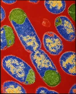
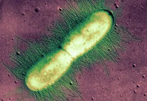
E. coli bacterium. Coloured transmission electron micrograph (TEM) of an Escherichia coli bacterium in the early stages of binary fission, the process by which the bacterium divides

Background imagePathogenic Collection: Cholera bacteria, artwork

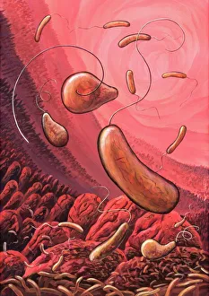
Cholera bacteria, artwork
Cholera bacteria. Artwork of Vibrio cholerae bacteria in the small intestine. These Gram-negative rod-shaped bacteria have a single polar flagellum (tail-like structure)

Background imagePathogenic Collection: Bacteria infecting a macrophage, SEM

Bacteria infecting a macrophage, SEM
Bacteria infecting a macrophage. Coloured scanning electron micrograph (SEM) of Mycobacterium tuberculosis bacteria (purple) infecting a macrophage white blood cell

Background imagePathogenic Collection: Pseudomonas aeruginosa bacteria, SEM

Pseudomonas aeruginosa bacteria, SEM
Pseudomonas aeruginosa bacteria, coloured scanning electron micrograph (SEM). These Gram-negative rod-shaped bacteria are found in soil and water, and as normal flora in the human intestine

Background imagePathogenic Collection: Adenovirus, artwork

Adenovirus, artwork
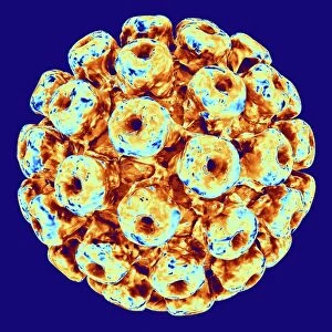
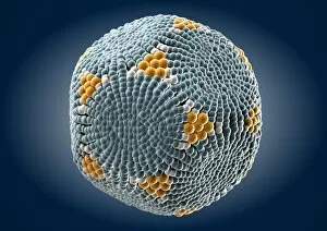
Adenovirus. Computer artwork of an adenovirus, showing the surface structure of the viruss outer protein coat (capsid). Adenoviruses are known to infect humans

Background imagePathogenic Collection: Polyoma BK virus, artwork C013 / 7465

Polyoma BK virus, artwork C013 / 7465
Computer artwork of the capsid of a polyoma BK virus. This polyomavirus is common in the urinary tract of adults, where it lives without harming its host

Background imagePathogenic Collection: Salmonella culture

Salmonella culture
MODEL RELEASED. Salmonella culture. Microbiologist holding a petri dish containing a culture of Salmonella sp. bacteria. These bacteria are the cause of salmonellosis (food poisoning) in humans

Background imagePathogenic Collection: Mouse malaria parasite, SEM

Mouse malaria parasite, SEM
Mouse malaria parasite. Coloured scanning electron micrograph (SEM) of a Plasmodium berghei protozoan (yellow) and red blood cells. P. berghei is the parasite that causes malaria in mice

Background imagePathogenic Collection: Rift Valley fever virus, TEM

Rift Valley fever virus, TEM

Background imagePathogenic Collection: Bladder infection

Bladder infection
Bacterial infection of bladder (cystitis). Colour Scanning Electron Micrograph (SEM) of the human bladder showing bacterial infection

Background imagePathogenic Collection: Paramyxovirus particles, TEM

Paramyxovirus particles, TEM
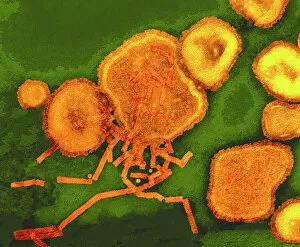
Sendai virus. Coloured transmission electron micrograph (TEM) of Sendai virus particles (virions, orange). The protein coat (capsid) of one of the particles has split

Background imagePathogenic Collection: E. coli 0157: H7 bacteria

E. coli 0157: H7 bacteria
E. coli 0157:H7 bacteria. Coloured scanning electron micrograph (SEM) of Escherichia coli 0157:H7 bacteria, cause of foodborne illness

Background imagePathogenic Collection: Rhinovirus and antibody, molecular model C015 / 7139

Rhinovirus and antibody, molecular model C015 / 7139
Rhinovirus. Molecular model of the antigen-binding fragment (Fab) from a strongly neutralising antibody bound to a human rhinovirus 14 (HRV-14) particle

Background imagePathogenic Collection: Phagocytosis of fungus spores, SEM

Phagocytosis of fungus spores, SEM
Phagocytosis of fungus spores. Coloured scanning electron micrograph (SEM) of spores from the fungus Aspergillus fumigatus (red) being phagocytosed (engulfed and destroyed) by a dendritic cell (brown)

Background imagePathogenic Collection: Giardia lamblia protozoa, micrograph

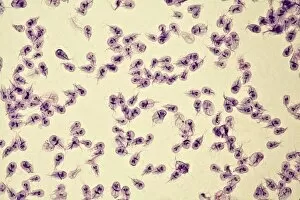
Giardia lamblia protozoa, micrograph
Giardia lamblia protozoa, light micrograph. These single-celled parasitic protozoa infect the intestinal tract and are most common in tropical regions

Background imagePathogenic Collection: HIV infected macrophage, SEM C018 / 8598

HIV infected macrophage, SEM C018 / 8598
HIV infected macrophage. Coloured ion-abrasion scanning electron micrograph (IA-SEM) of a macrophage white blood cell infected with human immunodeficiency virus (HIV, red)

Background imagePathogenic Collection: White blood cell antigen presentation C016 / 9058

White blood cell antigen presentation C016 / 9058
White blood cell antigen presentation. Coloured scanning electron micrograph (SEM) showing the interaction between a macrophage (yellow) and a T helper lymphocyte (Th cell, green)

Background imagePathogenic Collection: Helicobacter pylori bacteria, SEM C016 / 9136

Helicobacter pylori bacteria, SEM C016 / 9136
Helicobacter pylori bacteria. Coloured scanning electron micrograph (SEM) of Helicobacter pylori bacteria (green) in the stomach. H

Background imagePathogenic Collection: Helicobacter pylori bacteria, SEM C016 / 9053

Helicobacter pylori bacteria, SEM C016 / 9053
Helicobacter pylori bacteria. Coloured scanning electron micrograph (SEM) of a Helicobacter pylori bacterium (blue) in the stomach. H

Background imagePathogenic Collection: Hepatitis B virus particles, artwork C016 / 9097

Hepatitis B virus particles, artwork C016 / 9097
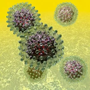
Hepatitis B virus (HBV) particles, computer artwork. Each particle consists of an outer lipid envelope (green) that surrounds a protein nucleopcapsid (purple)

Background imagePathogenic Collection: Rhinovirus and antibody, molecular model C015 / 7138

Rhinovirus and antibody, molecular model C015 / 7138
Rhinovirus. Molecular model of the antigen-binding fragment (Fab) from a strongly neutralising antibody bound to a human rhinovirus 14 (HRV-14) particle

Background imagePathogenic Collection: Influenza virus particles, TEM

Influenza virus particles, TEM
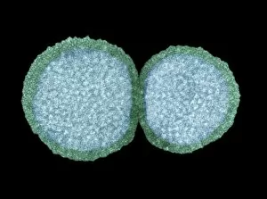
Influenza virus particles, coloured transmission electron micrograph (TEM). The virus consists of ribonucleic acid (RNA), surrounded by a nucleocapsid (blue) and a lipid envelope (green)

Background imagePathogenic Collection: Parvovirus particle, artwork C013 / 4640

Parvovirus particle, artwork C013 / 4640
Parvovirus particle. Computer artwork showing the outer structure of a human parvovirus (family Parvoviridae) particle (virion)

Background imagePathogenic Collection: Viral recognition by antibodies, artwork C013 / 4722

Viral recognition by antibodies, artwork C013 / 4722
Viral recognition by antibodies. Computer artwork of rabies (family Rhabdoviridae) virus particles (virions, pink) being identified by monoclonal antibodies (Y-shaped, blue)

Background imagePathogenic Collection: Ducks and bird flu virus particles

Ducks and bird flu virus particles
Ducks and avian influenza virus particles, composite image. The virus particles (brown) have been imaged using a transmission electron microscope (TEM)

Background imagePathogenic Collection: HIV viruses, TEM

HIV viruses, TEM
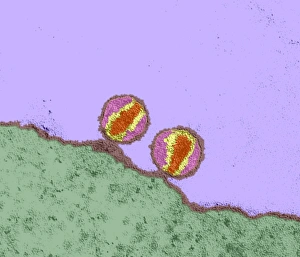
HIV viruses. Coloured transmission electron micrograph (TEM) of human immunodeficiency virus (HIV) particles. These particles are docking at a T-lymphocyte (white blood cell) just before infection

Background imagePathogenic Collection: Bacteria research

Bacteria research
MODEL RELEASED. Bacteria research. Researcher holding an empty petri dish in front of other dishes containing cultured bacteria (dots)

Background imagePathogenic Collection: Smallpox virus particle, TEM

Smallpox virus particle, TEM
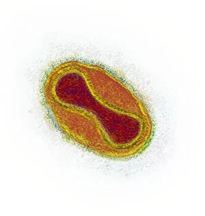
Smallpox virus particle. Coloured transmission electron micrograph (TEM) of a Variola major virus particle. Each particle consists of a DNA (deoxyribonucleic acid)

Background imagePathogenic Collection: Infectious bronchitis virus (IBV), TEM

Infectious bronchitis virus (IBV), TEM
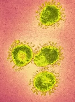
Infectious bronchitis virus (IBV) particles, coloured transmission electron micrograph (TEM). This virus is a member of the coronavirus family

Background imagePathogenic Collection: Curvularia geniculata fungus, SEM

Curvularia geniculata fungus, SEM
Curvularia geniculata fungus. Coloured scanning electron micrograph (SEM) of the fruiting body of the mould fungus Curvularia geniculata

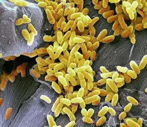
Background imagePathogenic Collection: Bacillus subtilis bacteria, SEM

Bacillus subtilis bacteria, SEM
Bacillus subtilis. Coloured scanning electron micrograph (SEM) of Bacillus subtilis bacteria. B. subtilis, or hay bacillus, is an aerobic, gram-positive bacillus(rod-shaped bacterium)

Background imagePathogenic Collection: Bacteria on lung hairs

Bacteria on lung hairs
Bacteria in a lung. Computer artwork of bacteria (yellow) on cilia (hair-like structures) in a lung. Bacterial infection of the lungs can cause potentially fatal pneumonia

Background imagePathogenic Collection: Spirochete bacteria, TEM

Spirochete bacteria, TEM
Spirochete bacteria, coloured transmission electron micrograph (TEM). Spirochetes are bacteria with helically coiled cells

Background imagePathogenic Collection: Rod-shaped bacteria

Rod-shaped bacteria. Computer artwork of rod- shaped bacteria (bacilli). Typical bacilli are E. coli and Salmonella bacteria, though there are many others as well

Background imagePathogenic Collection: Yeast cells, TEM

Yeast cells, TEM
Yeast cells, coloured transmission electron micrograph (TEM). The cell wall is shown in blue, cytoplasm in red, and vacuole in green. Yeast is a single-celled fungus that reproduces asexually

Background imagePathogenic Collection: Plague bacteria

Plague bacteria. Light micrograph of Gram- stained Yersinia pestis bacteria. This bacterium is the cause of bubonic plague (generally agreed to have been the Black Death of the Middle Ages)

Background imagePathogenic Collection: Fungal spores, SEM

Fungal spores, SEM
Fungal spores. Coloured scanning electron micrograph (SEM) of fungal spores on a fruiting body of an Aspergillus sp. fungus. The name for such a fruiting body is a conidiophore

Background imagePathogenic Collection: Schistosome fluke, SEM

Schistosome fluke, SEM
Schistosome fluke. Coloured scanning electron micrograph (SEM) of a schistosome (Schistosoma sp.) fluke worm, a cause of schistosomiasis in humans

Background imagePathogenic Collection: Eurasian Badger (Meles meles) bovine tuberculosis vaccination scheme

Eurasian Badger (Meles meles) bovine tuberculosis vaccination scheme, badger in live trap being vaccinated by Wildlife Trust personnel, Shropshire, England, June

Background imagePathogenic Collection: Haemophilus, artwork F007 / 6739

Haemophilus, artwork F007 / 6739
Haemophilus bacteria, computer artwork

Background imagePathogenic Collection: MRSA bacteria, artwork F007 / 7431

MRSA bacteria, artwork F007 / 7431
MRSA (methicillin-resistant Staphylococcus aureus) bacteria, computer artwork

Background imagePathogenic Collection: Bacillus bacteria, artwork F007 / 6735

Bacillus bacteria, artwork F007 / 6735
Bacillus bacteria, computer artwork

Background imagePathogenic Collection: Coccus bacteria, artwork F007 / 6761

Coccus bacteria, artwork F007 / 6761
Coccus bacteria, Computer artwork

Background imagePathogenic Collection: Propionibacterium acnes, artwork F007 / 6754

Propionibacterium acnes, artwork F007 / 6754
Propionibacterium acnes bacteria, computer artwork

Background imagePathogenic Collection: Chlamydia, artwork F007 / 6763

Chlamydia, artwork F007 / 6763
Chlamydia bacteria, computer artwork

Background imagePathogenic Collection: Chlamydia, artwork F007 / 6746

Chlamydia, artwork F007 / 6746
Chlamydia bacteria, computer artwork

Background imagePathogenic Collection: Streptococcus, artwork F007 / 6760

Streptococcus, artwork F007 / 6760
Streptococcus bacteria, computer artwork

Background imagePathogenic Collection: Clostridium, artwork F007 / 6757

Clostridium, artwork F007 / 6757
Clostridium bacteria, computer artwork

Background imagePathogenic Collection: Peptostreptococcus, artwork F007 / 6736

Peptostreptococcus, artwork F007 / 6736
Peptostreptococcus bacteria, computer artwork

Background imagePathogenic Collection: Klebsiella, artwork F007 / 6742

Klebsiella, artwork F007 / 6742
Klebsiella bacteria, computer artwork

Background imagePathogenic Collection: Haemophilus, artwork F007 / 6764

Haemophilus, artwork F007 / 6764
Haemophilus bacteria, computer artwork

Background imagePathogenic Collection: Pseudomonas, artwork F007 / 6762

Pseudomonas, artwork F007 / 6762
Pseudomonas bacteria, computer artwork

Background imagePathogenic Collection: Mycoplasma pneumoniae, artwork F007 / 6755

Mycoplasma pneumoniae, artwork F007 / 6755
Mycoplasma pneumoniae bacteria, computer artwork

Background imagePathogenic Collection: Acinetobacter, artwork F007 / 6749

Acinetobacter, artwork F007 / 6749
Acinetobacter bacteria, computer artwork

Background imagePathogenic Collection: Streptococcus, artwork F007 / 6743

Streptococcus, artwork F007 / 6743
Streptococcus bacteria, computer artwork

Background imagePathogenic Collection: Peptostreptococcus, artwork F007 / 6753

Peptostreptococcus, artwork F007 / 6753
Peptostreptococcus bacteria, computer artwork

Background imagePathogenic Collection: Coccus bacteria, artwork F007 / 6744

Coccus bacteria, artwork F007 / 6744
Coccus bacteria, Computer artwork

Background imagePathogenic Collection: Bacillus bacteria, artwork F007 / 6752

Bacillus bacteria, artwork F007 / 6752
Bacillus bacteria, computer artwork

Background imagePathogenic Collection: Haemophilus, artwork F007 / 6756

Haemophilus, artwork F007 / 6756
Haemophilus bacteria, computer artwork


All Professionally Made to Order for Quick Shipping

Pathogenic microorganisms are microscopic entities that pose a significant threat to human health. From budding yeast cells to avian flu viruses, these tiny organisms have the potential to cause widespread diseases and infections. In the world of pathogens, the avian flu virus stands out as one of the most notorious culprits. Its ability to jump from birds to humans has sparked global concerns about pandemics and led scientists on a quest for effective prevention strategies. Neutrophils, our body's first line of defense against infection, play a crucial role in engulfing harmful bacteria like MRSA (methicillin-resistant Staphylococcus aureus). This SEM image captures this incredible process, showcasing how neutrophils work tirelessly to protect us from pathogenic invaders. Salmonella bacteria are another well-known pathogen responsible for causing foodborne illnesses worldwide. This SEM image provides an up-close look at their unique structure and highlights their capacity for survival in various environments. The HIV particle is infamous for its devastating impact on the immune system. Understanding its intricate structure has been instrumental in developing antiretroviral therapies that help manage this deadly virus. Anthrax cultures depicted in historical diagrams remind us of past outbreaks and highlight the importance of ongoing research into preventing bioterrorism threats associated with this highly infectious disease. Norovirus particles captured through TEM imaging reveal their distinctive shape and shed light on why they are so contagious. Responsible for many cases of gastroenteritis, noroviruses can spread rapidly within communities if proper hygiene measures aren't followed diligently. Tuberculosis bacteria continue to be a major global health concern due to their ability to infect millions each year. Efforts towards improved diagnostics and treatment options remain critical in combating this persistent pathogen. E. coli bacteria serve as both harmless inhabitants of our gut flora but also dangerous pathogens when certain strains acquire virulence factors. This diverse bacterium reminds us that not all E. coli strains are created equal.

© Copyright Media Storehouse, All Rights Reserved 2003 - 2024
Facebook     Pinterest     100% Payment Secure     Reviews IO