
Palp Collection
"Exploring the Intricate World of Palps: Tick Mouthparts and SEM Revealing Fascinating Details" In this captivating journey, we delve into the mesmerizing realm of palps


All Professionally Made to Order for Quick Shipping
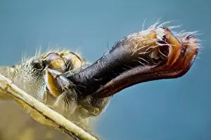
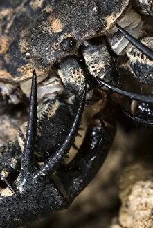
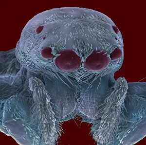
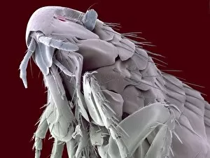
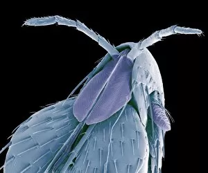
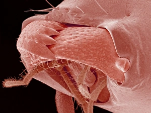
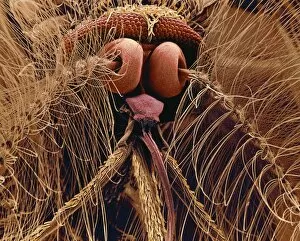
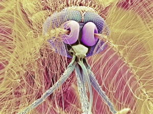
"Exploring the Intricate World of Palps: Tick Mouthparts and SEM Revealing Fascinating Details" In this captivating journey, we delve into the mesmerizing realm of palps. These tiny structures, often overlooked, hold remarkable secrets waiting to be unraveled. Through scanning electron microscopy (SEM), we are granted an up-close look at their intricate beauty. We begin with a close-up of the head and palps of a Giant House Spider (Tegenaria gigantea) adult male. The delicate tick mouthparts and sensory organs come alive under the lens, showcasing nature's precision craftsmanship. Moving on, we encounter a Nursery web spider gracefully carrying an egg sac. Its palp acts as both guide and protector for its precious cargo—a testament to the incredible adaptability found in arachnids. The world of jumping spiders is next on our exploration agenda. Their vibrant colors and unique features make them irresistible subjects for study. With SEM technology, we witness their expressive faces up close—their eyes gleaming with curiosity and determination. A Harvestman Palp Terminal captured through SEM reveals astonishing details that might have otherwise gone unnoticed by human eyes alone. This glimpse into their world highlights just how much there is still left to discover about these enigmatic creatures. Our adventure takes us further into Central Africa where we encounter the Variegated Tailless Whip Scorpion—an intriguing arachnid known for its fearsome appearance but gentle nature. Close-ups reveal not only its cephalothorax with piercing eyes but also its formidable pincers ready to defend against any threat that may arise. Finally, another peek at a Jumping Spider's head through SEM reminds us once again of their undeniable charm—tiny yet mighty predators equipped with exceptional vision and agility. Through these glimpses into the lives of various arachnids, we gain appreciation for the complexity hidden within even seemingly small organisms like palps.