Fraud Blocker Skip to main content

Orthopoxvirus Collection

Orthopoxvirus, a microscopic entity that has left an indelible mark on human history

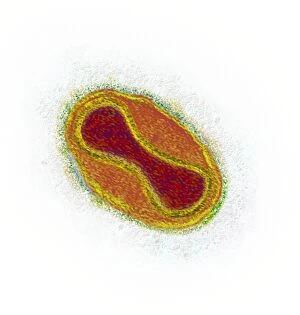
Background imageOrthopoxvirus Collection: Smallpox virus particle, TEM

Smallpox virus particle, TEM
Smallpox virus particle. Coloured transmission electron micrograph (TEM) of a Variola major virus particle. Each particle consists of a DNA (deoxyribonucleic acid)

Background imageOrthopoxvirus Collection: Microscopic view of samllpox

Microscopic view of samllpox

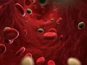
Background imageOrthopoxvirus Collection: Smallpox infection, artwork

Smallpox infection, artwork
Smallpox infection. Computer artwork of smallpox virus particles in the bloodstream

Background imageOrthopoxvirus Collection: Monkeypox virus particles, TEM C016 / 7386

Monkeypox virus particles, TEM C016 / 7386
Monkeypox virus (MPV) particles, coloured transmission electron micrograph (TEM). Each particle is composed of a DNA (deoxyribonucleic acid) genome surrounded by a protein coat and lipid envelope

Background imageOrthopoxvirus Collection: Monkeypox virus particles, TEM C016 / 7387

Monkeypox virus particles, TEM C016 / 7387
Monkeypox virus (MPV) particles, coloured transmission electron micrograph (TEM). Each particle is composed of a DNA (deoxyribonucleic acid) genome surrounded by a protein coat and lipid envelope

Background imageOrthopoxvirus Collection: Monkeypox virus particles, TEM C016 / 7385

Monkeypox virus particles, TEM C016 / 7385
Monkeypox virus (MPV) particles, coloured transmission electron micrograph (TEM). Each particle is composed of a DNA (deoxyribonucleic acid) genome surrounded by a protein coat and lipid envelope

Background imageOrthopoxvirus Collection: Monkeypox virus particles, TEM C016 / 7384

Monkeypox virus particles, TEM C016 / 7384
Monkeypox virus (MPV) particles, coloured transmission electron micrograph (TEM). Each particle is composed of a DNA (deoxyribonucleic acid) genome surrounded by a protein coat and lipid envelope

Background imageOrthopoxvirus Collection: Monkeypox virus particle, TEM C016 / 7383

Monkeypox virus particle, TEM C016 / 7383
Monkeypox virus (MPV) particle, coloured transmission electron micrograph (TEM). Each particle is composed of a DNA (deoxyribonucleic acid) genome surrounded by a protein coat and lipid envelope

Background imageOrthopoxvirus Collection: Monkeypox virus particle, TEM C016 / 7380

Monkeypox virus particle, TEM C016 / 7380
Monkeypox virus (MPV) particle, coloured transmission electron micrograph (TEM). Each particle is composed of a DNA (deoxyribonucleic acid) genome surrounded by a protein coat and lipid envelope

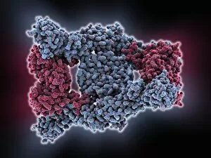
Background imageOrthopoxvirus Collection: Interferon antagonism by viral protein C015 / 5421

Interferon antagonism by viral protein C015 / 5421
Interferon (IFN) antagonism by viral protein. Molecular model of an orthopoxvirus IFN-gamma-binding protein tetramer bound to an IFN-gamma dimer

Background imageOrthopoxvirus Collection: Interferon antagonism by viral protein C015 / 5420

Interferon antagonism by viral protein C015 / 5420
Interferon (IFN) antagonism by viral protein. Molecular model of an orthopoxvirus IFN-gamma-binding protein tetramer bound to an IFN-gamma dimer

Background imageOrthopoxvirus Collection: Smallpox variola viruses

Smallpox variola viruses
Smallpox virus. Coloured transmission electron micrograph (TEM) of Orthopoxvirus variola viruses, the causative agent of smallpox

Background imageOrthopoxvirus Collection: Vaccinia virus particles, SEM

Vaccinia virus particles, SEM
Vaccinia virus particles. Coloured scanning electron micrograph (SEM) of vaccinia virus particles (red) budding from a cell (blue and green) after replicating

Background imageOrthopoxvirus Collection: Vaccinia viruses, TEM

Vaccinia viruses, TEM
Vaccinia virus particles. Coloured transmission electron micrograph (TEM) of sectioned vaccinia virus particles. The genetic cores (red) are covered by membrane layers (green)

Background imageOrthopoxvirus Collection: Vaccinia virus particles, TEM

Vaccinia virus particles, TEM


All Professionally Made to Order for Quick Shipping

Orthopoxvirus, a microscopic entity that has left an indelible mark on human history. Its most notorious representative, smallpox, is captured here in all its terrifying glory. The microscopic view of smallpox reveals the intricate structure and complexity of this ancient enemy. Smallpox infection, depicted through artwork, serves as a haunting reminder of the devastation caused by this virus throughout centuries. It spread like wildfire, leaving countless lives shattered in its wake. But orthopoxviruses don't stop at smallpox alone; they also encompass monkeypox. These tiny particles can be observed under the powerful lens of a transmission electron microscope (TEM). Monkeypox virus particles appear both fascinating and menacing simultaneously. The TEM images reveal the distinct features of monkeypox viruses - their shape and arrangement hinting at their potential to wreak havoc within living organisms. Each particle carries with it the ability to disrupt normal cellular function and trigger disease. Interestingly, these viruses possess proteins capable of antagonizing interferon - our body's natural defense mechanism against viral infections. The image showcasing interferon antagonism by viral protein sheds light on how orthopoxviruses cunningly evade our immune system's attempts to eliminate them. Orthopoxvirus may have plagued humanity for centuries but thanks to advancements in science and medicine, we now live in a world free from smallpox due to successful vaccination campaigns. However, vigilance remains crucial as emerging threats like monkeypox remind us that these ancient adversaries still persist in nature. As we continue our battle against infectious diseases, understanding orthopoviruses becomes paramount for developing effective prevention strategies and treatments. Through scientific exploration and collaboration across borders, we strive towards eradicating not just one specific virus but ensuring global health security for generations to come.