
Organism Collection (page 5)
"Unveiling the Diversity of Organisms: From Ancient Avian Dinosaurs to Microscopic Marvels" In this captivating journey through time and scale


All Professionally Made to Order for Quick Shipping
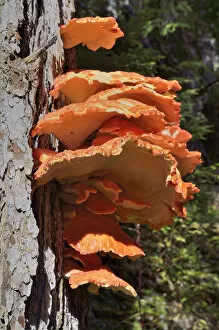
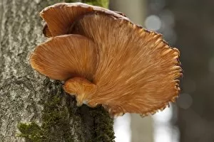
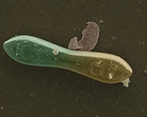
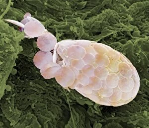
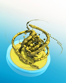
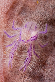
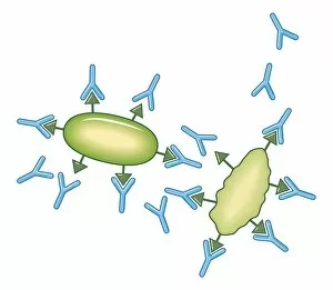
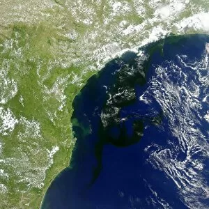
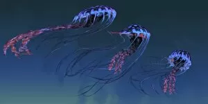
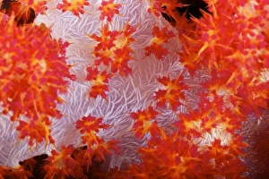
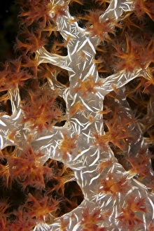
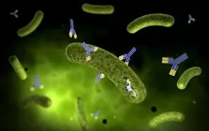
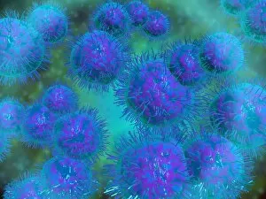
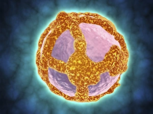
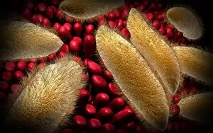
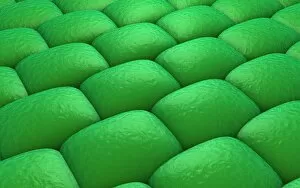
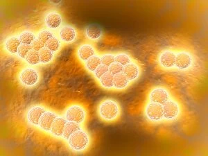
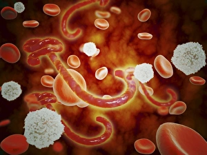
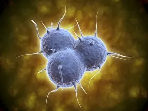
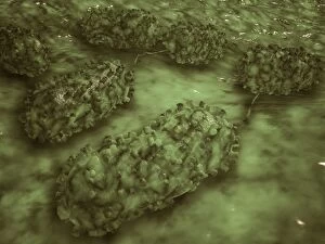
"Unveiling the Diversity of Organisms: From Ancient Avian Dinosaurs to Microscopic Marvels" In this captivating journey through time and scale, we explore the fascinating world of organisms. Our first stop takes us back millions of years, where we encounter Archaeopteryx, a bird-like dinosaur that once soared across prehistoric skies. Picture No. 11675590 allows us a glimpse into its remarkable existence. Moving forward in history, Aspidonia greets us with its historical artwork from 1899. This masterpiece showcases the intricate beauty of an organism yet unknown to many. Palaeontology, c1910 introduces us to another mysterious creature whose creator remains anonymous but leaves behind invaluable knowledge for future generations. Shifting our focus towards microscopic life forms, we delve into the realm of Protozoa – single-celled organisms that scavenge for particles and microorganisms or absorb nutrients from their environment. Witnessing their survival strategies is truly awe-inspiring. Microscopic views grant us access to two notorious organisms - human respiratory syncytial virus and chlamydia - both causing significant health concerns worldwide. These images serve as reminders of the complex interplay between humans and these tiny entities. Venturing underwater reveals breathtaking wonders such as sea fan X-ray imagery showcasing vibrant marine life thriving amidst coral reefs' splendorous ecosystem. Diatoms captured through scanning electron microscopy offer a glimpse into nature's artistic side while highlighting their crucial role in aquatic habitats. Our exploration wouldn't be complete without acknowledging scientific advancements in understanding diseases like syphilis; projection slides from the 1920s provide insight into this infamous organism's structure and impact on human health. As our journey nears its end, let's take a moment to appreciate nature's harmony by witnessing a fly gracefully soaring over a mesmerizing reef landscape—a reminder that even within vast ecosystems teeming with diverse organisms, every individual plays an essential role in maintaining the delicate balance of life.