Fraud Blocker Skip to main content

Oral Collection (page 5)

"Exploring the Intricacies of Oral Health

Background imageOral Collection: Gingivitis, SEM C013 / 7135

Gingivitis, SEM C013 / 7135
Gingivitis. Coloured scanning electron micrograph (SEM) of a large number of bacteria (yellow) in the gingiva (gums) of a human mouth

Background imageOral Collection: Gingivitis, SEM C013 / 7134

Gingivitis, SEM C013 / 7134
Gingivitis. Coloured scanning electron micrograph (SEM) of a large number of bacteria (green) in the gingiva (gums) of a human mouth

Background imageOral Collection: Oral statement by the American Navy admiral

Oral statement by the American Navy admiral. Japanese print shows three men, two seated and one standing, possibly Commander Anan, age 54; Perry, age 49; Captain Henry Adams, age 59

Background imageOral Collection: Coral polyp mouth

Coral polyp mouth. Close-up of the mouth (pink, centre) of a hard coral polyp (class Anthozoa). This marine invertebrate uses its tentacles (not clearly seen) to catch plankton from the water

Background imageOral Collection: Mouse tooth, SEM

Mouse tooth, SEM
Mouse tooth. Coloured scanning electron micrograph (SEM) of a molar from a mouse (Mus musculus). The tooth is similar to a human tooth but has more pronounced cusps (projections on the upper surface)

Background imageOral Collection: Boy shouting into a megaphone

Boy shouting into a megaphone

Background imageOral Collection: Mouse molars, SEM

Mouse molars, SEM
Mouse molars. Coloured scanning electron micrograph (SEM) of mouse (Mus musculus) molar teeth. The teeth are very similar to human teeth

Background imageOral Collection: Human tongue

Human tongue
MODEL RELEASED. Tongue of a young teenager showing prominent taste buds

Background imageOral Collection: Pierced tongue

Pierced tongue
MODEL RELEASED. Pierced tongue. Woman with a metal stud through her tongue. This type of piercing has come in for criticism from dentists, who believe that it can cause infections

Background imageOral Collection: Mouse incisor, SEM

Mouse incisor, SEM
Mouse incisor. Coloured scanning electron micrograph (SEM) of a mouse (Mus musculus) incisor that has been left to grow. This tooth is comprised mainly of dentin

Background imageOral Collection: Set of teeth

Set of teeth, computer artwork

Background imageOral Collection: Tin can telephone

Tin can telephone being used by two boys. The boy at left is speaking into one of the cans, and the sound waves are being transmitted along the string to the other can

Background imageOral Collection: Tooth decay, SEM

Tooth decay, SEM
Tooth decay. Coloured scanning electron micrograph (SEM) of dental plaque (brown) on a human incisor. Plaque is a film of bacteria embedded in a glycoprotein matrix

Background imageOral Collection: Gold tooth

Gold tooth. Computer artwork of a set of adult teeth, where one of the incisors has been replaced with a gold tooth. Teeth may need to be replaced due to decay or injury

Background imageOral Collection: Coloured SEM of a cut dental inlay

Coloured SEM of a cut dental inlay
Dental inlay. (Image 3 of 3). Coloured scanning electron micrograph (SEM) of a dental inlay (blue) on a mount (brown). The inlay has just come from the miniature milling machine in which it was cut

Background imageOral Collection: Dental pin, SEM

Dental pin, SEM
Dental pin. Coloured scanning electron micrograph (SEM) of a dental pin. This type of pin is used to keep false teeth in place

Background imageOral Collection: Used and unused toothpicks, SEM

Used and unused toothpicks, SEM
Used and unused toothpicks. Coloured scanning electron micrograph (SEM) of an unused (left) and a used (right) toothpicks. The used toothpick has a blunt end and is covered in dental plaque

Background imageOral Collection: X-ray of teeth

X-ray of teeth
False-colour dental x-ray of teeth from the right hand side of a 45 year old male mouth. The red areas are fillings, while the green area in the tooth at bottom left is a patch of caries

Background imageOral Collection: Used toothbrush bristle, SEM

Used toothbrush bristle, SEM
Used toothbrush bristle. Coloured scanning electron micrograph (SEM) of dental plaque on a bristle from a used toothbrush. Plaque consists of a film of bacteria embedded in a glycoprotein matrix

Background imageOral Collection: Extracted molar

Extracted molar
Macrophotograph of an extracted molar (male, upper, number 7), showing the build up of tartar, formed from salts in the saliva, around the gum line (brown line). Magnification: X1.2 (at 35mm size)

Background imageOral Collection: Coloured X-ray of impacted wisdom tooth and brace

Coloured X-ray of impacted wisdom tooth and brace
Impacted wisdom tooth. Coloured X-ray of the mouth showing an impacted wisdom tooth (highlighted yellow/magenta, centre left)

Background imageOral Collection: Coloured SEM of a glass fibre dental ceramic

Coloured SEM of a glass fibre dental ceramic
Glass fibre dental ceramic. Coloured scanning electron micrograph (SEM) of a dental ceramic reinforced with glass fibres. This ceramic is used to make artificial teeth such as those used in dental

Background imageOral Collection: Polarised LM of a tooth with a dental crown

Polarised LM of a tooth with a dental crown
Dental crown. Polarised light micrograph (PLM) of a longitudinal section through a tooth (yellow & blue) with a dental crown (grainy area)

Background imageOral Collection: Gum disease

Gum disease. Computer artwork of diseased gums and teeth. Gum disease causes inflammation and bleeding of the gums. It is the most common cause of tooth loss

Background imageOral Collection: Coloured SEM of a dental drill drilling into tooth

Coloured SEM of a dental drill drilling into tooth
Dental drill on tooth. Coloured scanning electron micrograph (SEM) of a diamond-tipped dental drill (orange) drilling a hole into a tooth (blue)

Background imageOral Collection: Coloured SEM of a tooth with a dental crown

Coloured SEM of a tooth with a dental crown
Dental crown. Coloured scanning electron micrograph (SEM) of a tooth (green) with a dental crown (light blue). The tooth and ceramic crown are coated in debris (orange)

Background imageOral Collection: Dental X-ray

Dental X-ray
MODEL RELEASED. Dental X-ray. Dentist looking at an X-ray of a patients teeth. This is a panoramic wraparound X-ray that shows all the teeth of both the upper and lower jaw

Background imageOral Collection: Coloured SEM of the join between a tooth and crown

Coloured SEM of the join between a tooth and crown
Dental crown. Coloured scanning electron microgr- aph (SEM) of the join between a tooth (green) and a dental crown (light blue)

Background imageOral Collection: Artwork of tooth showing periodontal disease

Artwork of tooth showing periodontal disease
Periodontal disease. Artwork of a section through a human tooth with periodontal disease of its surrounding gum and bone. The tooths white enamel covers the dentine (yellow) and pulp (purple)

Background imageOral Collection: Dental drill, SEM

Dental drill, SEM
Dental drill. Coloured scanning electron micrograph (SEM) of the tip of a dental drill bit. Magnification: x28 when printed at 10 centimetres wide

Background imageOral Collection: Decay on molar tooth, SEM

Decay on molar tooth, SEM
Tooth decay. Scanning electron micrograph (SEM) of the surface of a decaying molar tooth. The surface enamel (white) has begun to break down (for example, at lower right)

Background imageOral Collection: Dental inlay being cut in a milling machine

Dental inlay being cut in a milling machine

Background imageOral Collection: Extracted molar showing 3 amalgam fillings

Extracted molar showing 3 amalgam fillings
Macrophotograph of an extracted molar (male, upper, number 7), a top view showing three 20 year old amalgam fillings. The filling is a powdered alloy consisting of silver, tin

Background imageOral Collection: Computer graphic of a tooth ready for dental inlay

Computer graphic of a tooth ready for dental inlay
Dental inlay preparation. (Image 1 of 3). Computer graphic of a tooth with a cavity cut into it in preparation for a dental inlay

Background imageOral Collection: Coloured SEM of the root of a dental implant

Coloured SEM of the root of a dental implant

Background imageOral Collection: Dental plaque

Dental plaque
Tooth plaque. Dental plaque (yellow) on a patients teeth. Plaque is the main cause of tooth decay. It is a sticky coating that consists of saliva, bacteria and particles of food

Background imageOral Collection: Pearl between teeth

Pearl between teeth. Conceptual image of a pearl held between a womans teeth. This could represent teeth that are clean, healthy, white, polished or " pearly"

Background imageOral Collection: Polarised LM of a cracked ceramic dental crown

Polarised LM of a cracked ceramic dental crown
Dental crown. Polarised light micrograph (PLM) of a longitudinal section through a cracked dental crown made of metal (multicoloured) and ceramic (above metal)

Background imageOral Collection: Polarised LM of a tooth with various fillings

Polarised LM of a tooth with various fillings
Tooth fillings. Polarised light micrograph (PLM) of a longitudinal section through a tooth with various fillings. The enamel (orange) has been replaced in areas of decay (dental caries)

Background imageOral Collection: Trapped food in dental braces

Trapped food in dental braces
MODEL RELEASED. Trapped food in dental braces, which are fixed to the teeth of a teenage girl. Fixed braces consist of metal brackets cemented to each tooth

Background imageOral Collection: Young girl with fever has oral temperature taken

Young girl with fever has oral temperature taken
MODEL RELEASED. Measuring oral temperature. Feverish young girl with a thermometer in her mouth looks up at her mother. After a few minutes the thermometer will register the correct temperature

Background imageOral Collection: Rabbit tongues surface, SEM

Rabbit tongues surface, SEM
Rabbit tongues surface, coloured scanning electron micrograph (SEM). The tongue is covered in thin projections called filiform papillae. These sense touch

Background imageOral Collection: Tongue papillae, light micrograph

Tongue papillae, light micrograph
Tongue papillae. Coloured light micrograph of a section through the tongue, showing its filiform papillae (red). They project from the tongues upper surface, giving it its rough texture

Background imageOral Collection: Taste bud, light micrograph

Taste bud, light micrograph
Taste bud. Coloured light micrograph of a section through the tongue, showing a taste bud. This taste bud is seen as a round purple mass slightly to the right of the vertical white line

Background imageOral Collection: Blood vessels from the gums, SEM

Blood vessels from the gums, SEM
Blood vessels from the gums, coloured scanning electron micrograph (SEM). This is a cast: the vessels were filled with a resin, which then set

Background imageOral Collection: Toothbrush

Toothbrush, computer artwork

Background imageOral Collection: Dentist and patient

Dentist and patient. Conceptual artwork of a patient warding off a dentist with toothpaste. The dentist is holding a dental mirror at upper left and a dental probe at upper right

Background imageOral Collection: View from inside the mouth of dental crown fitting

View from inside the mouth of dental crown fitting


All Professionally Made to Order for Quick Shipping

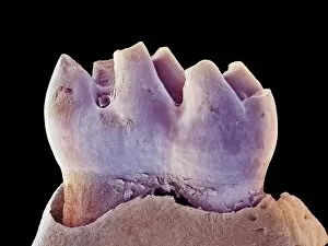
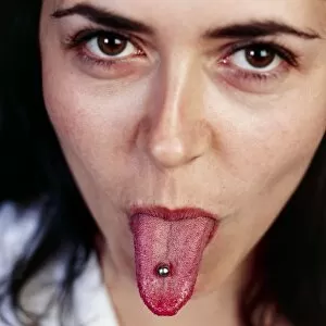
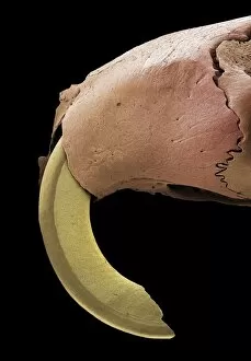
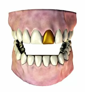
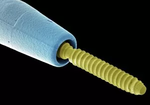
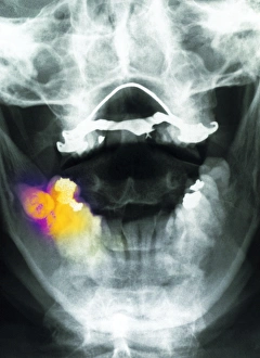
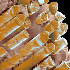
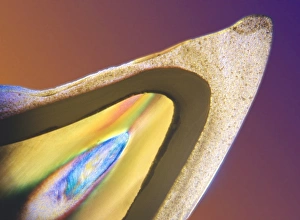
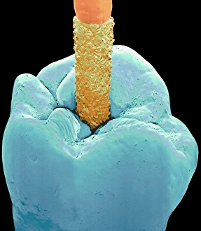
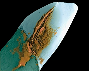
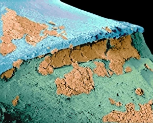
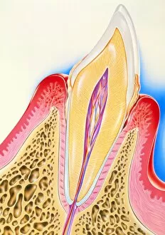
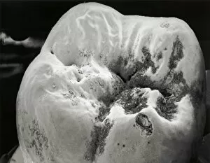
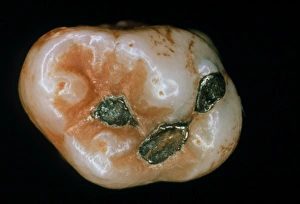
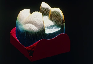
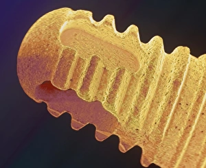
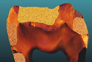
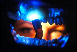
"Exploring the Intricacies of Oral Health: From Ancient Art to Modern Science" Step into the world health as we unravel its fascinating secrets through a diverse range of captivating images. Leonardo da Vinci's meticulous "Skull anatomy" drawing takes us back in time, showcasing his unparalleled understanding of dental structures. A panoramic dental X-ray reveals a comprehensive view, allowing dentists to diagnose and treat conditions with precision. Meanwhile, in Senegal, a Griot skillfully plays the Kora, reminding us of the cultural significance attached to oral traditions and storytelling. False teeth serve as a testament to mankind's quest for restoring smiles and functionality. A polarised LM image showcases decay within a molar tooth, highlighting the importance of regular check-ups and proper oral hygiene practices. The intricate illustration depicting our respiratory system reminds us that our oral cavity is not isolated but intricately connected to our overall well-being. Understanding this connection helps prevent various diseases affecting both our mouth and lungs. Delving deeper into dental knowledge, an illustration unveils the complex anatomy behind our teeth – each playing an essential role in chewing and speech production. Used dental floss under SEM magnification emphasizes the significance of interdental cleaning for maintaining optimal oral health. An X-ray revealing a fractured jawbone serves as a reminder that accidents can impact not only aesthetics but also functional aspects of one's smile. However, advancements like Januvia diabetes drug molecule offer hope by addressing underlying conditions affecting oral health. Zooming closer into modern dentistry marvels, an LM cross-section showcases how dental implants seamlessly integrate with surrounding tissues – revolutionizing tooth replacement options for countless individuals worldwide. Lastly, we witness everyday care routines as a man diligently brushes his teeth – reinforcing that simple habits lay the foundation for lifelong oral wellness. In this journey through artistry and science intertwined with human experiences across cultures and time periods, it becomes evident that caring for your mouth goes beyond just a smile – it is an essential part of overall health and well-being.