
Optic Nerves Collection
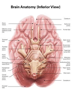
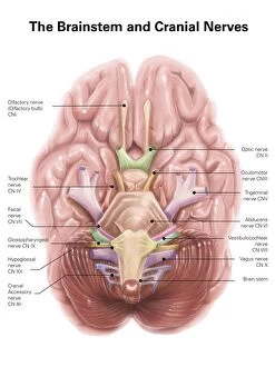
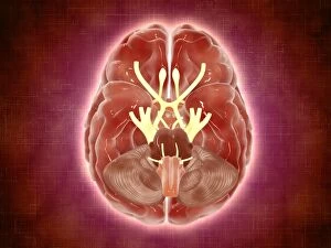
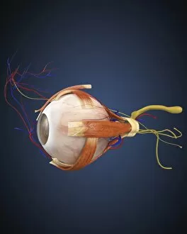
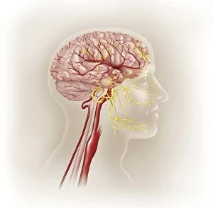
The optic nerves, an intricate part of the anatomy of the human brain, can be seen from an inferior view


All Professionally Made to Order for Quick Shipping
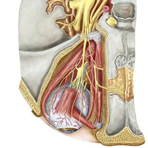
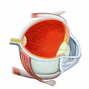
The optic nerves, an intricate part of the anatomy of the human brain, can be seen from an inferior view. These anatomical pathways are responsible for innervating the lacrimal gland, ensuring our eyes stay moist and healthy. In a conceptual image depicting human eye anatomy, we get a glimpse into the complex structure that allows us to see. The eye is intricately connected to the skull, working together seamlessly to provide us with vision. However, not all images of the human eye show perfect health. In one such image, we witness macular degeneration affecting the retina. This condition highlights how crucial they can in transmitting visual information from our eyes to our brain. Switching perspectives to explore orbital cuts reveals fascinating details about these nerves' connections within our skulls. Here we find the abducent nerve alongside ciliary ganglion and oculomotor nerve - all playing vital roles in controlling eye movements and focusing abilities. Returning to conceptual images of human eye anatomy showcases both normal retinas and those affected by advanced diabetic retinopathy. It serves as a reminder that they can vulnerable and require proper care for optimal functioning. Understanding these complexities helps us appreciate how remarkable our visual system truly is. The optic nerves serve as conduits between our eyes and brains, allowing us to perceive and interpret the world around us through sight – an extraordinary gift indeed.