
Occlusion Collection
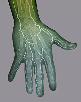
"Unlocking the Mystery of Occlusion: Exploring the Intricate Web of Blocked Blood Vessels" Ischaemia and Digital Angiogram


All Professionally Made to Order for Quick Shipping
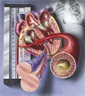
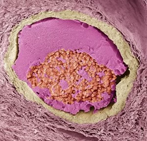
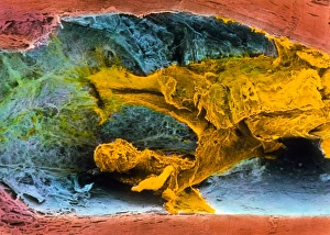
"Unlocking the Mystery of Occlusion: Exploring the Intricate Web of Blocked Blood Vessels" Ischaemia and Digital Angiogram: Peering into the Restricted Flow Thrombosed Blood Vessel meets Artwork C013 / 4649: A Visual Representation of Blockage Cholesterol vs Plaque in Arteries: The Battle for a Healthy Heart Unveiling Atherosclerotic Plaque in Heart Arteries: An Alarming Discovery Good vs Bad Cholesterol in the Bloodstream: Balancing Health Risks Acute Coronary Syndrome and Microvascular Obstruction: When Occlusion Turns Critical Interior View of Heart Reveals Muscle Cells and Atherosclerotic Artery Detail Thrombus Formation on Valve within Vein - A Closer Look at Clotting Mechanisms Platelets, White Blood Cells, and Red Blood Cells Coexist Within a Blocked Blood Vessel Normalcy vs Chaos - Comparing Healthy Arteries to Plaque & Thrombus Formation Artery Exposed.