
Occluded Collection
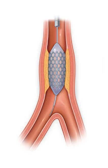
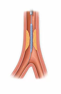
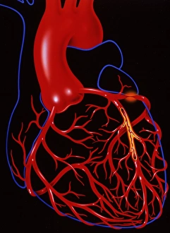
"Unveiling the Hidden Dangers: Exploring the World Vessels" A glimpse into the intricate world of cardiovascular health reveals a vessel occluded with plaque


All Professionally Made to Order for Quick Shipping
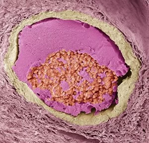
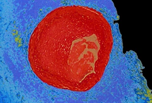
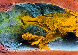
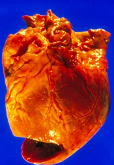
"Unveiling the Hidden Dangers: Exploring the World Vessels" A glimpse into the intricate world of cardiovascular health reveals a vessel occluded with plaque, showcasing a stent in place and balloon inflated – an innovative solution to restore blood flow. Witnessing the impact of plaque buildup, we observe a vessel occluded with plaque where a stent has been strategically placed, offering hope for improved circulation and cardiac function. In the realm of medical artistry, we encounter a striking depiction of a thrombosed blood vessel (artwork C013/4649), reminding us of the delicate balance between life and potential blockages. Peering through the lens of scientific exploration, we are captivated by an up-close view of a blood clot (SEM C016/9088) - nature's way of alerting us to potential dangers lurking within our veins. Continuing our journey into microscopic landscapes, another captivating image emerges - revealing yet another intricately formed blood clot (SEM C016/9089), emphasizing the importance of early detection and prevention. Delving deeper into coronary complications, we uncover an astonishing SEM image showcasing a coronary blood clot (SEM C016/9133), serving as both an educational tool and reminder to prioritize heart health. Shifting gears towards diagnostic imaging techniques, X-ray images reveal large bowel obstructions that demand immediate attention for effective treatment (X-ray C017/7855 & X-ray C017/7854). With clarity provided by modern technology, an X-ray highlights a blocked artery - urging individuals to be proactive in maintaining their vascular well-being while seeking timely medical intervention when necessary. Through vibrant colors under light microscopy comes forth an artistic representation depicting an occluded human coronary artery – inviting awe at both its beauty and significance in understanding cardiovascular diseases.