
Occipital Lobe Collection
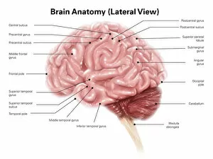
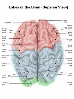
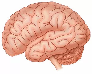
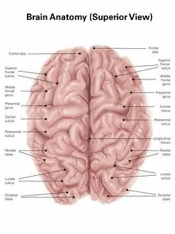
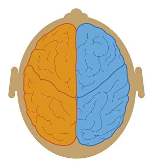
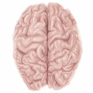
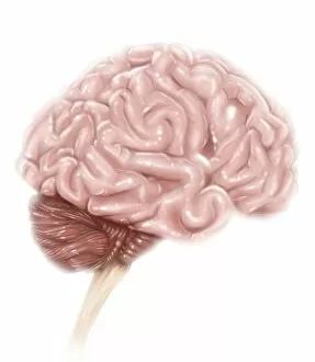
The occipital lobe, located at the back of the brain, plays a crucial role in our visual perception


All Professionally Made to Order for Quick Shipping
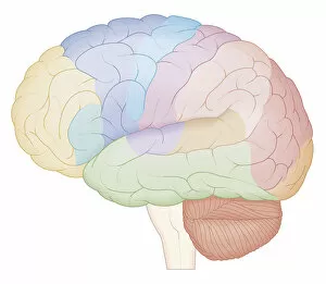
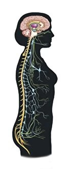
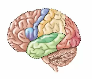
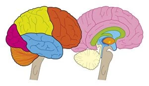
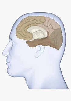
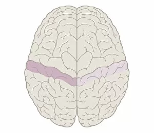
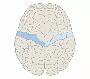
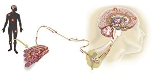
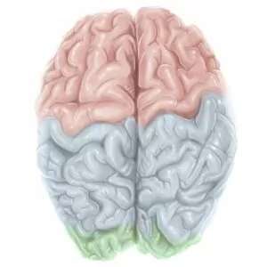
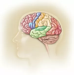
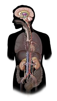
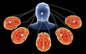
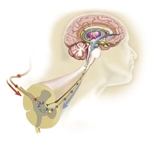
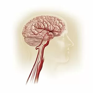
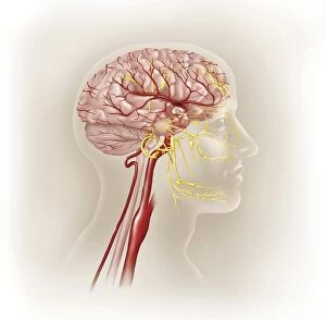
The occipital lobe, located at the back of the brain, plays a crucial role in our visual perception. In this superior view of the human brain with colored lobes and labels, we can clearly identify this important region. As we shift to a lateral view of a normal brain, it becomes evident how the occipital lobe interacts with other structures such as the basal ganglia. Examining an intricate artwork showcasing the basal ganglia further emphasizes its connection to this vital sensory processing center. Meanwhile, arteries of The Head remind us of the intricate network that supplies blood to every part of our brain, including the occipital lobe. In this superior view illustration depicting human brain anatomy, we gain insight into how different regions work together harmoniously. A cross-section biomedical illustration provides a detailed map highlighting various areas within our brains - including where we find the occipital lobe. Moving beyond just neurological aspects, another image reveals both autonomic nervous system and limbic system within our body. This reminds us that while vision is processed in the occipital lobe, emotions and involuntary bodily functions are also interconnected here. A side view displaying functional lobes allows us to appreciate how each area serves distinct purposes; among them lies our trusty occipital lobe responsible for transforming light into meaningful images. Finally, a digital illustration showcases not only these lobes but also offers an insightful cross-section perspective on their inner workings. To round out our exploration beyond just cerebral matters: neck vertebra anatomy engraving from 1866 demonstrates how intricately connected all parts of our body are - even those seemingly unrelated like bones in your neck. Similarly enlightening is frontal trunk anatomy engraving from 1866 which highlights connections between organs and systems throughout your entire being.