
Occipital Bone Collection
The occipital bone, located at the base of the human skull, is a vital component that plays a crucial role in protecting our brain and supporting various functions


All Professionally Made to Order for Quick Shipping
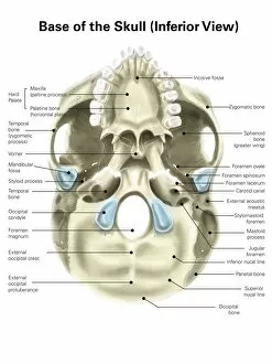
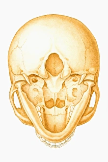
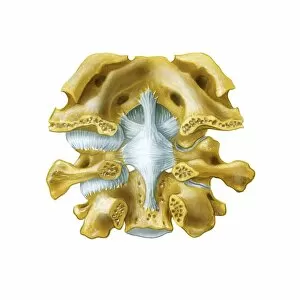
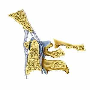
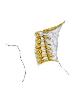
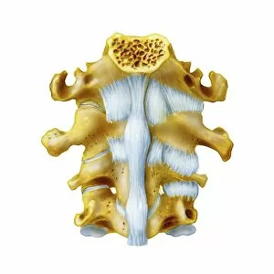
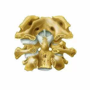
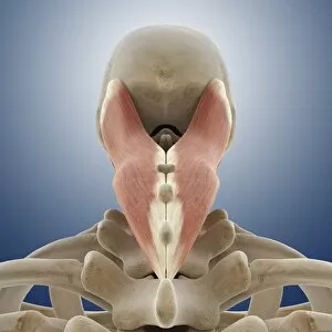
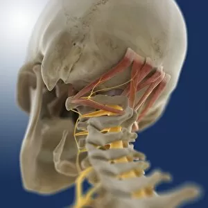
The occipital bone, located at the base of the human skull, is a vital component that plays a crucial role in protecting our brain and supporting various functions. In this captivating image, we are presented with an inferior view of the colored base of the human skull, meticulously labeled to enhance our understanding. Accompanying this visual masterpiece is another artwork showcasing the intricate suboccipital muscles and nerve. The attention to detail in this piece allows us to appreciate the complexity and interconnectedness of these structures. As we delve deeper into anatomical engravings from 1866, we are treated to a glimpse of back torso anatomy. These engravings provide us with valuable insights into how different components within our body work together seamlessly. Moving on, an illustration captures our attention as it presents a unique perspective - a view of the human skull seen from below. This depiction not only showcases the beauty and intricacy of our skeletal structure but also highlights how each element fits perfectly like pieces in a puzzle. Returning to colored images, we explore further details regarding back muscles. The artwork provides clarity on their placement and function within our musculoskeletal system. Lastly, we encounter artworks depicting important joints related to the occipital bone: atlanto-occipital joint and vertebral joints of the neck. These illustrations shed light on how these joints facilitate movement while maintaining stability in one's neck region. Through these captivating visuals ranging from engravings dating back centuries ago to modern-day illustrations with labels, we gain profound knowledge about one integral part -the occipital bone- which forms an essential foundation for numerous bodily functions.