Fraud Blocker Skip to main content

Neurotransmitter Collection (#3)

"Exploring the Intricacies of Neurotransmitters

Background imageNeurotransmitter Collection: Dopamine receptor D3 C016 / 4449

Dopamine receptor D3 C016 / 4449
D(3) dopamine receptor is a protein that in humans is encoded by the DRD3 gene.This gene encodes the D3 subtype of the dopamine receptor

Background imageNeurotransmitter Collection: Neuropeptide Y neurotransmitter molecule C014 / 0013

Neuropeptide Y neurotransmitter molecule C014 / 0013
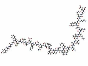
Neuropeptide Y neurotransmitter molecule. Molecular model showing the structure of the neurotransmitter neuropeptide Y (NPY)

Background imageNeurotransmitter Collection: Neural network, artwork C013 / 4636

Neural network, artwork C013 / 4636
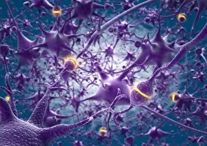
Neural network. Computer artwork of nerve cells (neurons) connected by processes (filaments), known as dendrites and axons, to form a neural network

Background imageNeurotransmitter Collection: Neuromuscular synapse, light micrograph

Neuromuscular synapse, light micrograph
Neuromuscular junction. Fluorescent confocal light micrograph of the junction between a nerve cell and a muscle (not seen). The axon of the nerve cell (neuron) has been tagged with a blue dye

Background imageNeurotransmitter Collection: Artwork of a nerve cell of the brain & a synapse

Artwork of a nerve cell of the brain & a synapse
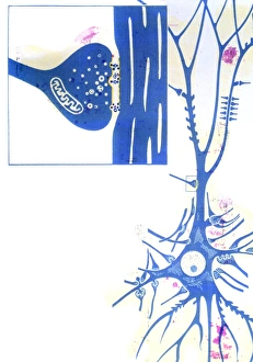
Nerve cell and synapses. Artwork of a nerve cell (neurone) of the brain with a close-up of a synapse. The nerve cell body is at lower right

Background imageNeurotransmitter Collection: Coloured TEM of a nerve synapse

Coloured TEM of a nerve synapse

Background imageNeurotransmitter Collection: Astrocyte / neuron synapse in the brain

Astrocyte / neuron synapse in the brain
Astrocyte/neuron synapse in the brain. Artwork of a nerve cell (neuron, yellow) synapse in the brain with an associated regulatory astrocyte cell (red)

Background imageNeurotransmitter Collection: Computer artwork of a nerve synapse

Computer artwork of a nerve synapse
Nerve synapse. Computer artwork of a nerve synapse. The synapse is a junction between a terminal (light blue) of one nerve cell (neurone) and another cell (green/purple)

Background imageNeurotransmitter Collection: Artwork of a nerve synapse

Artwork of a nerve synapse
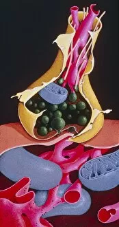
Synapse. Illustration of a synapse, the junction between two nerve cells. The green structures in the terminal swelling (bouton) of the pre-synaptic cell are synaptic vesicles

Background imageNeurotransmitter Collection: Action of a beta blocker drug, artwork

Action of a beta blocker drug, artwork
Action of a beta blocker drug on nerve synapses, artwork. Beta blockers are used to treat cardiac arrhythmia (abnormal heartbeats) and hypertension (high blood pressure)

Background imageNeurotransmitter Collection: Artwork showing sleeping drug action with face

Artwork showing sleeping drug action with face
Action of sleeping drugs. Artwork of the face of a sleeping woman cut away to show a magnified image of sleeping drugs acting on nerve cells (neurones, upper left & right) in her brain

Background imageNeurotransmitter Collection: Beta-endorphin molecule

Beta-endorphin molecule
Beta-endorphin, molecular model. This neurotransmitter causes insensitivity to pain and a feeling of well being. Atoms are represented as spheres and are colour-coded: carbon (grey)

Background imageNeurotransmitter Collection: Dopamine, 3D molecular model

Dopamine, 3D molecular model
3D molecular model of dopamine. Dopamine is a catecholamine neurotransmitter produced in different aereas of the brain. Dopamine is also a neurohormone released by the hypothalamus

Background imageNeurotransmitter Collection: Serotonin crystals, light micrograph

Serotonin crystals, light micrograph
Serotonin. Polarised light micrograph of serotonin, a neurotransmitter. Serotonin is derived from tryptophan, and is found in the vertebrate brain

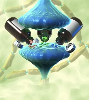
Background imageNeurotransmitter Collection: Neurological drug treatment, artwork

Neurological drug treatment, artwork
Neurological drug treatment, conceptual image. Computer artwork representing the use of drugs that affect neural networks by altering the transmission of neurotransmitters across synapses (blue)

Background imageNeurotransmitter Collection: Role of serotonin in the digestive system

Role of serotonin in the digestive system, artwork. In the gut serotonin (5HT) is produced by enterochromaffin (EC) cells (blue and white cells in upper circle)

Background imageNeurotransmitter Collection: Nerve synapse, artwork

Nerve synapse, artwork
Nerve synapse. Computer artwork of of a junction, or synapse, between two nerve cells (neurons). As the electrical signal reaches the presynaptic end of a neuron it triggers the release of

Background imageNeurotransmitter Collection: Synapse structure, artwork

Synapse structure, artwork
Synapse structure. Cutaway artwork showing the structure of a synapse, the point where two nerve ending meet. The electrical impulse moving along a nerve is transmitted to the adjacent nerve by

Background imageNeurotransmitter Collection: Synapse, artwork

Synapse, artwork
Synapse. Computer artwork of a synapse, the junction between nerve cells. Synapses transmit electrical signals from one nerve cell to the next

Background imageNeurotransmitter Collection: Synaptic endorphins, artwork

Synaptic endorphins, artwork
Synaptic endorphins. Artwork showing the release (top) of neurotransmitters called endorphins (yellow) from the end of a nerve (blue)

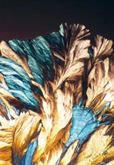
Background imageNeurotransmitter Collection: Adenosine crystals, light micrograph

Adenosine crystals, light micrograph
Adenosine crystals, polarised light micrograph. Adenosine is a nucleoside molecule composed of the base adenine bonded to the sugar ribose

Background imageNeurotransmitter Collection: Enkephalin crystals, light micrograph

Enkephalin crystals, light micrograph
Enkephalin crystals, polarised light micrograph. Enkephalin is an endorphin found in the human brain. There are two variants: Met-enkephalin (seen here), which contains the amino acid methionine

Background imageNeurotransmitter Collection: Intestinal endocrine cell, TEM

Intestinal endocrine cell, TEM
Intestinal endocrine cell, coloured transmission electron micrograph (TEM). This is a type 1 intestinal endocrine cell. The granules (red) contain serotonin

Background imageNeurotransmitter Collection: GABA crystals, light micrograph

GABA crystals, light micrograph
GABA crystals. Polarised light micrograph of gamma-aminobutyric acid (GABA). GABA is an inhibitory neurotransmitter (nerve signalling chemical) that regulates the excitability of all nerve cells

Background imageNeurotransmitter Collection: Acetylcholine crystals, light micrograph

Acetylcholine crystals, light micrograph
Acetylcholine crystals, polarised light micrograph. Acetylcholine (ACh) is a neurotransmitter (nerve signalling chemical) that plays an important role in relaying impulses at myoneural (muscle-nerve)

Background imageNeurotransmitter Collection: NMDA crystals, light micrograph

NMDA crystals, light micrograph
NMDA crystals, polarised light micrograph. NMDA (N-methyl-D-aspartic acid) is an amino acid derivative and a neurotransmitter (nerve signalling chemical)

Background imageNeurotransmitter Collection: Norepinephrine neurotransmitter molecule

Norepinephrine neurotransmitter molecule. Computer model showing the structure of the neurotransmitter and hormone norepinephrine, or noradrenaline

Background imageNeurotransmitter Collection: Met-enkephalin molecule

Met-enkephalin molecule. Computer model showing the structure of the neurotransmitter met-enkephalin (methionine-enkephalin)

Background imageNeurotransmitter Collection: Corticotropin-releasing factor complex

Corticotropin-releasing factor complex
Corticotropin-releasing factor (CRF) complex. Molecular model showing the structure of the human hormone and neurotransmitter CRF in complex with its receptor

Background imageNeurotransmitter Collection: Histamine molecule

Histamine molecule. Computer model showing the structure of a molecule of histamine. Atoms are colour-coded (carbon: dark grey, hydrogen: light grey, nitrogen: blue)

Background imageNeurotransmitter Collection: Glycine, molecular model

Glycine, molecular model
Glycine. Molecular model of the amino acid glycine. Its chemical formula is C2.H5.N.O2. Atoms are represented as balls and are colour- coded: carbon (blue), hydrogen (gold)

Background imageNeurotransmitter Collection: Aspartic acid, molecular model

Aspartic acid, molecular model
Aspartic acid. Molecular model of the amino acid aspartic acid. Its chemical formula is C4.H7.N.O4. Atoms are represented as balls and are colour- coded: carbon (blue), hydrogen (gold)

Background imageNeurotransmitter Collection: Glutamic acid, molecular model

Glutamic acid, molecular model
Glutamic acid. Molecular model of the amino acid glutamic acid. Its chemical formula is C5.H9.N.O4. Atoms are represented as rods and are colour- coded: carbon (blue), hydrogen (gold)

Background imageNeurotransmitter Collection: Cholinesterase enzyme

Cholinesterase enzyme. Molecular model of the secondary structure of butyrylcholinesterase (BChE), showing alpha helices (blue) and beta sheets (red and yellow)


All Professionally Made to Order for Quick Shipping

"Exploring the Intricacies of Neurotransmitters: Unveiling the Secrets of Brain Protein Research" The fascinating world of neurotransmitters unravels as scientists delve into brain protein research, seeking to understand the complex mechanisms that govern our thoughts and emotions. Intriguingly, cannabinoid receptor binding emerges as a crucial aspect in this intricate web. Artwork depicting these receptors highlights their role in modulating various physiological processes within our brains. As we journey deeper into the realm of neuroscience, we encounter schizophrenia—a disorder shrouded in mystery. Researchers tirelessly investigate how neurotransmitters contribute to its development and progression, hoping to unlock new treatment avenues for those affected. Amongst these chemical messengers lies oxytocin, known as the "love hormone. " Its unique structure is captured beautifully as a molecule, emphasizing its vital role in social bonding and trust. Nerve synapses become our next stop on this captivating voyage. Through TEM images, we witness the astonishing intricacy with which neurons communicate through electrical impulses and neurotransmitter release—bridging gaps between cells with astonishing precision. Serotonin takes center stage—an influential neurotransmitter responsible for regulating mood and sleep patterns. Visualizing it at a molecular level helps us appreciate its significance in maintaining emotional well-being. Enkephalin crystals shimmer under light micrographs—an opioid peptide offering insights into pain management strategies. Understanding their structures allows researchers to develop novel therapies targeting pain pathways effectively. Delving further into neurochemistry brings us face-to-face with ecstasy's impact on brain function—a topic both intriguing and concerning simultaneously. Scientists strive to comprehend how this recreational drug affects neurotransmission patterns while considering potential long-term consequences. Dopamine receptor D3 C016/4464 captures attention—a key player implicated in reward-motivated behavior and addiction disorders. Studying its structure provides valuable clues towards developing targeted interventions for substance abuse issues plaguing society today.