Fraud Blocker Skip to main content

Neurone Collection (page 4)

Neurons, the fundamental building blocks of our nervous system, are intricate and fascinating cells that enable communication within our bodies

Background imageNeurone Collection: Acupuncture, artwork

Acupuncture, artwork
Acupuncture. Computer artwork showing an acupuncture needle inserted into a patients skin and disrupting a nerve signal (red arrows)

Background imageNeurone Collection: Creutzfeldt-Jakob disease, MRI scan

Creutzfeldt-Jakob disease, MRI scan
Creutzfeldt-Jakob disease. Coloured magnetic resonance imaging (MRI) scan of an axial section through the brain of a 35-year-old patient

Background imageNeurone Collection: Myelinated nerve tissue, TEM

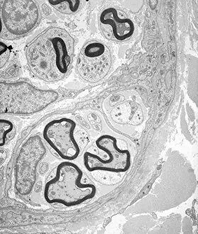
Myelinated nerve tissue, TEM
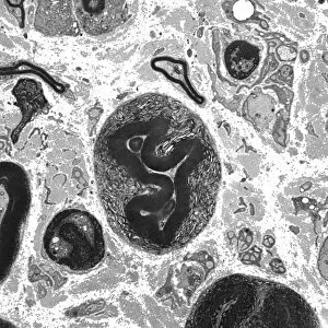
Myelinated nerve tissue. Transmission electron micrograph (TEM) of a section through myelinated nerve fibres (axons). Each axon is coated with many layers of myelin

Background imageNeurone Collection: Olfactory bulb anatomy, artwork

Olfactory bulb anatomy, artwork
Olfactory bulb anatomy. Artwork of a human nose, showing the anatomy of the olfactory bulb, the organ of smell. This lies above the anterior aspect of the nasal cavity on the ethmoid bone

Background imageNeurone Collection: Retina of the eye, light micrograph C016 / 0528

Retina of the eye, light micrograph C016 / 0528
Retina of the eye. Light micrograph of a section through the retina from a human eye. From top down: nerve fibres of the optic nerve and a blood vessel; several layers of neurons (nerve cells)

Background imageNeurone Collection: Nerve cell, illustration C018 / 0740

Nerve cell, illustration C018 / 0740
Nerve cell, or neuron, illustration. Neurons are responsible for passing information around the central nervous system (CNS) and from the CNS to the rest of the body

Background imageNeurone Collection: Cerebral cortex nerve cells C018 / 0062

Cerebral cortex nerve cells C018 / 0062
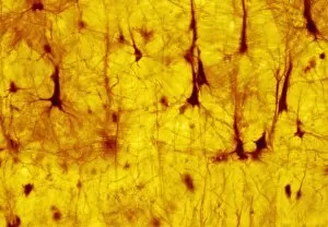
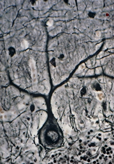
Cerebral cortex nerve cells. Light micrograph of a section through neurones (nerve cells, black) in the cerebral cortex of a human brain

Background imageNeurone Collection: Synapse, SEM C018 / 0122

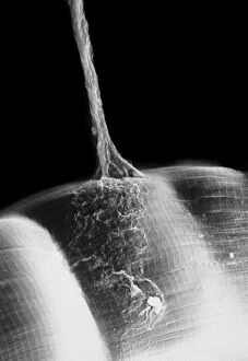
Synapse, SEM C018 / 0122
Synapse. Scanning electron micrograph (SEM) of a neuromuscular junction showing a motor neurone (vertical line) terminating on skeletal muscle fibres (across bottom frame)

Background imageNeurone Collection: Nerve synapse, artwork C017 / 3426

Nerve synapse, artwork C017 / 3426
Nerve synapse. Computer artwork of of a junction, or synapse, between two nerve cells (neurons). As the electrical signal reaches the presynaptic end of a neuron it triggers the release of

Background imageNeurone Collection: Myelin sheats and glial cells, artwork C014 / 2647

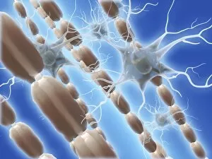
Myelin sheats and glial cells, artwork C014 / 2647
Computer artwork depicting axons surrounded by a myelin sheath (brown) and microglial cells (light blue). Glial cells are nervous system cells that provide structural support

Background imageNeurone Collection: Synapse, SEM

Synapse, SEM
Synapse. Coloured scanning electron micrograph (SEM) of a neuromuscular junction showing a motor neurone (green) terminating on skeletal muscle fibres (orange)

Background imageNeurone Collection: Induced nerve stem cells, micrograph

Induced nerve stem cells, micrograph
Induced nerve stem cells. Fluorescence light micrograph of neural (nerve) stem cells that have been created (induced) from human adult skin fibroblast cells by gene manipulation

Background imageNeurone Collection: Stem cell-derived nerve cells

Stem cell-derived nerve cells. Fluorescence light micrograph of neural (nerve) stem cells that have been derived from human embryonic stem cells (HESC)

Background imageNeurone Collection: Brain cells, artwork

Brain cells, artwork
Brain cells. Computer artwork representing neurons (nerve cells) in the brain

Background imageNeurone Collection: Brain nerve cells, TEM C014 / 0356

Brain nerve cells, TEM C014 / 0356
Brain nerve cells. Transmission electron micrograph (TEM) of a section through brain tissue from the cerebral cortex, showing numerous neurons (nerve cells) surrounded by axons and dendrites

Background imageNeurone Collection: Deep brain stimulation and nerve cell C016 / 7205

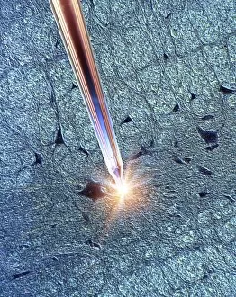
Deep brain stimulation and nerve cell C016 / 7205
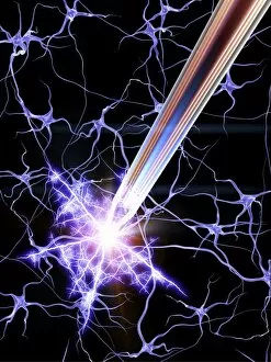
Deep brain stimulation. Computer artwork showing an electrode sending electrical impulses to nerve cells. In the background a light micrograph (LM) of a section through grey matter in the brain

Background imageNeurone Collection: Deep brain stimulation and nerve cell C016 / 7203

Deep brain stimulation and nerve cell C016 / 7203
Deep brain stimulation. Computer artwork showing an electrode sending electrical impulses to nerve cells. In the background a light micrograph (LM) of a section through grey matter in the brain

Background imageNeurone Collection: Deep brain stimulation and nerve cells C016 / 7206

Deep brain stimulation and nerve cells C016 / 7206
Deep brain stimulation (DBS). Computer artwork showing an electrode sending electrical impulses to nerve cells. DBS was developed for the treatment of Parkinsons disease

Background imageNeurone Collection: Purkinje nerve cell, TEM C014 / 0582

Purkinje nerve cell, TEM C014 / 0582
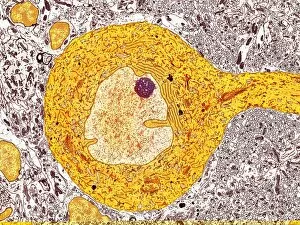
Purkinje nerve cell. Transmission electron micrograph (TEM) of a purkinje nerve cell (bright yellow, centre) from the cerebellum of the brain

Background imageNeurone Collection: Nerve cells, artwork C018 / 2887

Nerve cells, artwork C018 / 2887
Nerve cells, or neurons, computer artwork. Neurons are responsible for passing information around the central nervous system (CNS) and from the CNS to the rest of the body

Background imageNeurone Collection: Myelinated nerve, TEM C016 / 5840

Myelinated nerve, TEM C016 / 5840
Myelinated nerve. Coloured transmission electron micrograph (TEM) of a section through a myelinated nerve fibre and Schwann cell

Background imageNeurone Collection: Myelinated nerve, TEM C016 / 5839

Myelinated nerve, TEM C016 / 5839
Myelinated nerve. Coloured transmission electron micrograph (TEM) of a section through a myelinated nerve fibre and Schwann cell

Background imageNeurone Collection: Myelinated nerve, TEM C016 / 5838

Myelinated nerve, TEM C016 / 5838
Myelinated nerve. Coloured transmission electron micrograph (TEM) of a section through a myelinated nerve fibre and Schwann cell

Background imageNeurone Collection: Unmyelinated nerve, TEM C016 / 5805

Unmyelinated nerve, TEM C016 / 5805
Unmyelinated nerve. Transmission electron micrograph (TEM) of a section through axon (nerve fibre) bundles of unmyelinated nerves

Background imageNeurone Collection: Unmyelinated nerve, TEM C016 / 5804

Unmyelinated nerve, TEM C016 / 5804
Unmyelinated nerve. Transmission electron micrograph (TEM) of a section through axon (nerve fibre) bundles of unmyelinated nerves

Background imageNeurone Collection: Myelinated nerve, TEM C016 / 5448

Myelinated nerve, TEM C016 / 5448
Myelinated nerve. Transmission electron micrograph (TEM) of a section through a myelinated nerve fibre and Schwann cell. Myelin (black)

Background imageNeurone Collection: Myelinated nerve, TEM C016 / 5370

Myelinated nerve, TEM C016 / 5370
Myelinated nerve. Transmission electron micrograph (TEM) of a section through a myelinated nerve fibre and Schwann cell (centre)

Background imageNeurone Collection: Synapses, artwork C014 / 0002

Synapses, artwork C014 / 0002
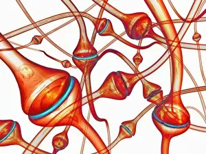
Synapses. Computer artwork of synapses, the junction between nerve cells (orange). Synapses transmit electrical signals from one nerve cell to the next

Background imageNeurone Collection: Synapses, artwork C014 / 0004

Synapses, artwork C014 / 0004
Synapses. Computer artwork of synapses, the junction between nerve cells (blue). Synapses transmit electrical signals from one nerve cell to the next

Background imageNeurone Collection: Synapses, artwork C014 / 0003

Synapses, artwork C014 / 0003
Synapses. Computer artwork of synapses, the junction between nerve cells (blue). Synapses transmit electrical signals from one nerve cell to the next

Background imageNeurone Collection: Nerve cell, conceptual artwork C013 / 9994

Nerve cell, conceptual artwork C013 / 9994
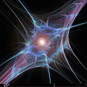
Nerve cell, conceptual computer artwork

Background imageNeurone Collection: Neural network, conceptual image C013 / 9958

Neural network, conceptual image C013 / 9958
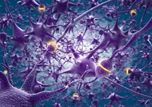
Neural network, conceptual image. Computer artwork representing interconnecting nerve cells (neurons)

Background imageNeurone Collection: Neural network, conceptual image C013 / 9957

Neural network, conceptual image C013 / 9957
Neural network, conceptual image. Computer artwork representing interconnecting nerve cells (neurons)

Background imageNeurone Collection: Nerve cell, SEM C013 / 9772

Nerve cell, SEM C013 / 9772
Nerve cell. Coloured scanning electron micrograph (SEM) of a nerve cell (neuron). Neurons are responsible for passing information around the central nervous system (CNS)

Background imageNeurone Collection: Cerebral cortex nerve cells C013 / 9767

Cerebral cortex nerve cells C013 / 9767
Cerebral cortex nerve cells. Light micrograph of a section through neurones (nerve cells, black) in the cerebral cortex of a human brain

Background imageNeurone Collection: Purkinje nerve cell C013 / 9763

Purkinje nerve cell C013 / 9763
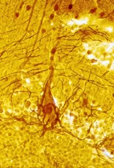
Purkinje nerve cell. Light micrograph of a purkinje nerve cell (orange, centre) from the cerebellum of the brain. The cell comprises a flask-shaped cell body from which numerous processes (dendrites)

Background imageNeurone Collection: Purkinje nerve cells C013 / 9745

Purkinje nerve cells C013 / 9745
Purkinje nerve cells. Light micrograph of three purkinje nerve cells (across bottom) from the cerebellum of the brain. The cells comprise a flask-shaped cell body from which numerous processes

Background imageNeurone Collection: Myelinated nerves, SEM C013 / 7142

Myelinated nerves, SEM C013 / 7142
Myelinated nerves. Coloured scanning electron micrograph (SEM) of a section through the sciatic nerve, showing the myelinated nerve fibres (axons)

Background imageNeurone Collection: Myelinated nerves, SEM C013 / 7141

Myelinated nerves, SEM C013 / 7141
Myelinated nerves. Coloured scanning electron micrograph (SEM) of a section through the sciatic nerve, showing the myelinated nerve fibres (axons)

Background imageNeurone Collection: Myelinated nerves, SEM C013 / 7138

Myelinated nerves, SEM C013 / 7138
Myelinated nerves. Coloured scanning electron micrograph (SEM) of a section through a myelinated nerve fibre (axon, beige, centre) from the sciatic nerve

Background imageNeurone Collection: Neural network, artwork C013 / 4636

Neural network, artwork C013 / 4636
Neural network. Computer artwork of nerve cells (neurons) connected by processes (filaments), known as dendrites and axons, to form a neural network

Background imageNeurone Collection: Retinal rod cell, TEM C013 / 4805

Retinal rod cell, TEM C013 / 4805
Retinal rod cell. Transmission electron micrograph (TEM) of a section through a rod cell from the retina of an eye, showing the inner segment (bottom) filled with mitochondria (green)

Background imageNeurone Collection: Retinal rod cell, TEM C013 / 4804

Retinal rod cell, TEM C013 / 4804
Retinal rod cell. Transmission electron micrograph (TEM) of a section through a rod cell from the retina of an eye, showing the inner segment (bottom) filled with mitochondria (green)

Background imageNeurone Collection: Nerve cell, TEM C013 / 4797

Nerve cell, TEM C013 / 4797
Nerve cell. Transmission electron micrograph (TEM) of a section through a neuron (nerve cell), showing characteristic Nissl body (dark blue lines), numerous golgi apparatus (curved green lines)

Background imageNeurone Collection: Nerve cell, TEM C013 / 4796

Nerve cell, TEM C013 / 4796
Nerve cell. Transmission electron micrograph (TEM) of a section through a neuron (nerve cell), showing characteristic Nissl body (dark red lines), numerous golgi apparatus (curved pink lines)

Background imageNeurone Collection: LM of a Purkinje cell in the cerebellum

LM of a Purkinje cell in the cerebellum

Background imageNeurone Collection: False-colour SEM of 3 neurones of cerebral cortex

False-colour SEM of 3 neurones of cerebral cortex
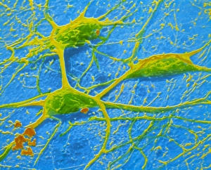
False-colour scanning electron micrograph (SEM) showing three neurones (nerve cells) of the human cerebral cortex - the outer grey matter of the brain

Background imageNeurone Collection: Brain nerve cells

Brain nerve cells
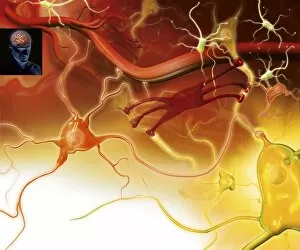
Nerve cells in the brain. Artwork showing the different types of nerve cells in the grey matter of the brain. Neurons (yellow, for example at lower right) relay nerve signals around the brain


All Professionally Made to Order for Quick Shipping

Neurons, the fundamental building blocks of our nervous system, are intricate and fascinating cells that enable communication within our bodies. This captivating image showcases nerve and glial cells under a light microscope, revealing their unique structures and functions. In another stunning capture, we witness the synapse nerve junction through a transmission electron microscope (TEM), where signals are transmitted between neurons. The complexity of this microscopic world is further highlighted in an immunofluorescent light micrograph depicting neurons and astrocytes, emphasizing their role in supporting neuronal function. A striking scanning electron microscope (SEM) image zooms in on a single nerve cell, showcasing its intricate details and delicate branching structure. These specialized cells play a crucial role in transmitting electrical impulses throughout our body's complex network. Moving beyond individual cells, we explore the vastness of glial stem cell culture under a light microscope. These versatile cells have the potential to differentiate into various types of support cells for neurons. The importance of blood supply to brain tissue is beautifully illustrated in an image capturing the cerebral cortex's intricate network of nerve cells nourished by tiny blood vessels. This symbiotic relationship ensures optimal functioning of our cognitive abilities. Delving deeper into neural stem cell culture reveals their remarkable ability to self-renew and differentiate into different types of brain cells. These incredible properties hold immense promise for regenerative medicine and understanding neurological disorders better. Lastly, we observe motor neurons under a light microscope—key players responsible for transmitting signals from the central nervous system to muscles or glands throughout our body. Their precise organization allows us to perform coordinated movements effortlessly. These captivating images provide glimpses into the mesmerizing world of neurones—a testament to nature's intricacy and beauty as they orchestrate every thought, sensation, movement within us while unraveling mysteries that continue to captivate scientists worldwide.