
Neuronal Collection
"Unveiling the Intricacies of Neuronal Wonders: From Nerve Cells to Ion Channels" In the vast realm of neuroscience, neuronal wonders never cease to amaze us


All Professionally Made to Order for Quick Shipping
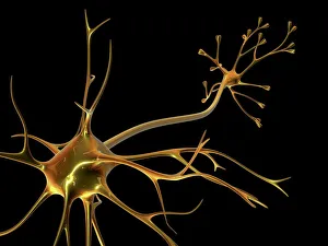
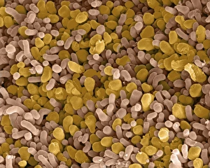
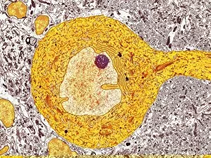
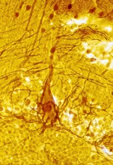
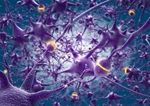
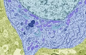
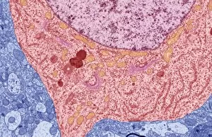
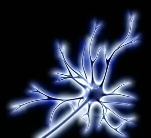
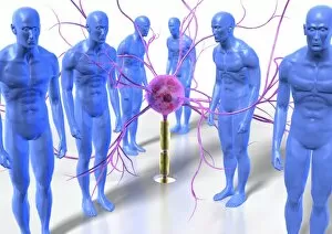
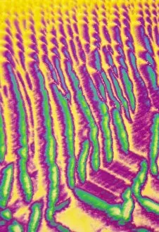
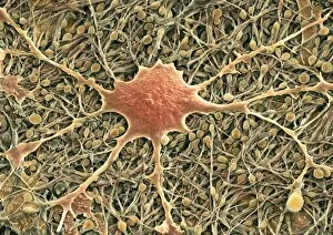
"Unveiling the Intricacies of Neuronal Wonders: From Nerve Cells to Ion Channels" In the vast realm of neuroscience, neuronal wonders never cease to amaze us. At the core of this intricate network lies the nerve cell, also known as a neuron. These remarkable cells transmit electrical signals throughout our bodies, allowing for communication between different parts. One captivating aspect is the rod and cone cells found in our eyes, responsible for vision. Under scanning electron microscopy (SEM), we witness their unique structures up close – rods aiding in low-light conditions and cones enabling color perception (SEM C014 / 4866). Delving deeper into neural architecture, we encounter Purkinje nerve cells that play a crucial role in coordinating movement and balance. Through transmission electron microscopy (TEM), these magnificent cells reveal their complex branching patterns (TEM C014 / 0583). Their significance cannot be overstated as they form vital connections within our cerebellum. Exploring further into neurochemistry, enkephalin crystals come into focus under light microscopy. These opioid peptides act as natural painkillers within our nervous system, providing relief when needed most (Light micrograph). Returning to visual marvels once more, SEM captures rod and cone cells with astonishing detail yet again (SEM C014 / 4864). The intricacy of these specialized neurons showcases nature's brilliance in crafting an organ capable of perceiving light so exquisitely. Zooming out from cellular structures brings us to KCNQ ion channel proteins – gatekeepers regulating electrical activity within neurons. Understanding their structure aids researchers in unraveling mysteries surrounding neurological disorders such as epilepsy or deafness (KCNQ ion channel protein structure). Venturing even deeper into molecular terrain reveals nitric oxide synthase molecules at work – key players involved in various physiological processes like blood pressure regulation and immune response modulation (F006 / 9452).