Fraud Blocker Skip to main content

Neurology Collection (page 64)

Neurology, the fascinating study of the nervous system, unravels the intricate workings of our brain and its various components

Background imageNeurology Collection: Cholinesterase enzyme

Cholinesterase enzyme. Molecular model of the secondary structure of butyrylcholinesterase (BChE), showing alpha helices (blue) and beta sheets (red and yellow)

Background imageNeurology Collection: Soman nerve agent molecule

Soman nerve agent molecule. Molecular model of Soman, also known as GD, a nerve agent used in chemical warfare. It acts as a neurotoxin by inhibiting cholinesterase enzymes needed for maintaining

Background imageNeurology Collection: Encephalin peptide

Encephalin peptide
Encephalin. Computer molecular graphic of part of a molecule of encephalin, a polypeptide found in the human brain. It has a painkilling effect

Background imageNeurology Collection: Cyclin-depenent kinase 5 molecule

Cyclin-depenent kinase 5 molecule
Cyclin-dependent kinase 5. Molecular model showing the secondary structure of the enzyme cylcin- dependent kinase 5 (cdk5)

Background imageNeurology Collection: VX nerve agent molecule

VX nerve agent molecule. Molecular model of VX, a nerve agent used in chemical warfare. VX blocks the action of an enzyme called acetylcholinesterase

Background imageNeurology Collection: Human brain, artwork

Human brain, artwork
Human brain anatomy. Computer artwork of a persons head showing the brain with the right hemisphere removed. The following regions can be seen: outer cortex (red) somatosensory cortex (lighter)

Background imageNeurology Collection: Human brain anatomy, artwork

Human brain anatomy, artwork
Human brain anatomy. Computer artwork of a persons head showing the brain with the right hemisphere removed. The brainstem can be seen (bottom left)

Background imageNeurology Collection: Cingulate gyrus in the brain, artwork

Cingulate gyrus in the brain, artwork
Temporal lobe in the brain. Computer artwork of a persons head showing the left hemisphere of the brain inside. The highlighted area shows the left temporal lobe

Background imageNeurology Collection: Cyborg brain, artwork

Cyborg brain, artwork
Cyborg brain, computer artwork. Computer artwork of a human brain with robotic implants emerging from the cerebellum and brainstem

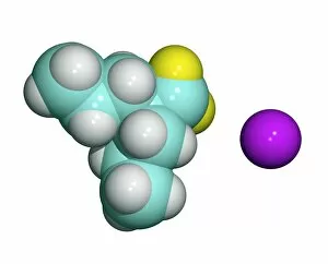
Background imageNeurology Collection: Sodium valproate, anti-epilepsy drug

Sodium valproate, anti-epilepsy drug
Sodium valproate drug, molecular model. This drug, marketed under dozens of brand names worldwide including Depacon and Epilim, is the sodium salt of valproic acid

Background imageNeurology Collection: Alprazolam drug molecule

Alprazolam drug molecule
Alprazolam. Computer model of a molecule of the drug alprazolam. It is sold under the brand name Xanax. It is a benzodiazepine, a sedative drug. It is sold by Pfizer

Background imageNeurology Collection: Zolpidem, sedative drug

Zolpidem, sedative drug
Zolpidem sedative drug, molecular model. This drug, marketed under several brand names including Ambien and Stilnoct, is used for the short-term treatment of insomnia

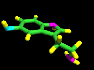
Background imageNeurology Collection: Serotonin neurotransmitter

Serotonin neurotransmitter
Serotonin. Computer model of a molecule of the neurotransmitter serotonin (5-hydroxytryptamine, C10.H12.N2.O). Atoms (tubes) are colour-coded: carbon (green), hydrogen (yellow)

Background imageNeurology Collection: Sumatriptan drug for migraine headaches

Sumatriptan drug for migraine headaches
Sumatriptan drug, molecular model. This drug, marketed as Imitrex, Imigran and Imigran Recovery, is used to treat migraine headaches

Background imageNeurology Collection: Cobra venom action, molecular model

Cobra venom action, molecular model
Cobra (Naja sp.) venom action. Molecular model showing top (upper centre) and side (lower centre) views of the secondary structure of an alpha-cobratoxin (snake venom protein)

Background imageNeurology Collection: Donepezil drug molecule

Donepezil drug molecule
Donepezil. Computer model of a molecule of the drug donepezil. It is sold under the brand name Aricept. It is an acetylcholinesterase inhibitor drug

Background imageNeurology Collection: Neotrofin drug molecule

Neotrofin drug molecule
Neotrofin. Computer model of a molecule of the drug known by the brand name Neotrofin, but generically known as leteprinim. It is also known as AIT-082

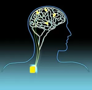
Background imageNeurology Collection: Parkinsons disease treatment, artwork

Parkinsons disease treatment, artwork
Parkinsons disease treatment. Computer artwork of electrodes, which have been surgically implanted in the brain to treat the tremors caused by Parkinsons disease

Background imageNeurology Collection: Pentetrazol drug molecule

Pentetrazol drug molecule. Computer model of Pentetrazol (PTZ), a potential drug candidate for Downs syndrome, formally used as a circulatory and respiratory stimulant under the trade name Metrazol

Background imageNeurology Collection: Zolpidem brain drug, molecular model

Zolpidem brain drug, molecular model
Zolpidem brain drug. Molecular model of the drug zolpidem (C19.H21.N3.O) with an artwork of the human brain. This drug is mainly used to treat insomnia

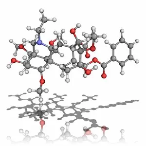
Background imageNeurology Collection: Aconite poison molecule

Aconite poison molecule. Molecular model of aconitine, a highly poisonous chemical found in aconite (Aconitum sp.) plants. Its chemical formula is C34H47NO11

Background imageNeurology Collection: Buspirone anti-anxiety drug molecule

Buspirone anti-anxiety drug molecule
Buspirone anti-anxiety drug. Computer model of a molecule of buspirone, marketed as Buspar, an anxiolytic (anti-anxiety) drug

Background imageNeurology Collection: Ampalex drug molecule

Ampalex drug molecule
Ampalex. Computer model of a molecule of the drug made under the brand name Ampalex. It is also known by its trial name of CX-516. It is an ampakine drug, used to treat Alzheimers disease

Background imageNeurology Collection: Tacrine drug molecule

Tacrine drug molecule
Tacrine. Computer model of a molecule of the drug tacrine. It is sold under the brand name Cognex. It is an acetylcholinesterase inhibitor drug

Background imageNeurology Collection: Dopamine neurotransmitter molecule

Dopamine neurotransmitter molecule
Dopamine molecule. Computer artwork of a molecule of the neurotransmitter and neurohormone dopamine (C8.H11.N.O2). The atoms are represented as spheres and are colour-coded: carbon (black)

Background imageNeurology Collection: Alpha-endorphin molecule

Alpha-endorphin molecule. Molecular model of the analgesic (painkilling) peptide alpha-endorphin. This molecule is released by the pituitary gland at times of stress or great pain

Background imageNeurology Collection: Spinal cord, transverse section

Spinal cord, transverse section
Spinal cord. Light micrograph of a transverse section through a human spinal cord. The spinal cord has a central canal around which are the dorsal and ventral horns (orange)

Background imageNeurology Collection: Alzheimers disease, artwork

Alzheimers disease, artwork
Alzheimers disease. Computer artwork of a vertical (coronal) slice through the brain of an Alzheimer patient (left) compared with a normal brain (right)

Background imageNeurology Collection: Nerve cell trauma response

Nerve cell trauma response. Fluorescent light micrograph of a section through a spinal cord affected by multiple sclerosis (MS)

Background imageNeurology Collection: Brain tissue, light micrograph

Brain tissue, light micrograph
Brain tissue. Light micrograph of a section through tissue from the mammalian brain, in this case from the cerebellum. The cerebellum is the part of the brain that controls sensory perception

Background imageNeurology Collection: Brain activity, artwork

Brain activity, artwork
Brain activity. Computer artwork showing activity (red) in the frontal (left) and temporal (centre) lobes of a human brain. The front of the brain is at left

Background imageNeurology Collection: Stroke, conceptual artwork

Stroke, conceptual artwork
Stroke. Conceptual computer artwork of a stroke, showing a cut-away of a human head and a blood vessel bursting in the brain. A stroke is also known as a cerebrovascular accident (CVA)


All Professionally Made to Order for Quick Shipping

Neurology, the fascinating study of the nervous system, unravels the intricate workings of our brain and its various components. From the Motor homunculus model that depicts how different body parts are represented in our motor cortex to the Histological Diagram of a Mammalian Retina revealing the complexity of our visual processing, every aspect is awe-inspiring. The Cerebellum tissue, as seen through a light micrograph, showcases its unique structure responsible for coordinating movement and balance. Meanwhile, an inferior view of the Anatomy of human brain highlights its remarkable organization and interconnectedness. Through Brain fibres captured by DTI MRI scans like C017 / 7099 or C017 / 7035, we gain insights into neural pathways that facilitate communication between different regions. These pathways form a complex network crucial for transmitting information throughout our brain. Exploring Brain blood vessels using a 3D angiogram (C007 / 1981) reveals their vital role in supplying oxygen and nutrients to sustain neuronal function. Additionally, understanding Motor and sensory homunculi helps us comprehend how specific areas within our brains control different bodily functions. Delving deeper into neurology brings us face-to-face with Medulla oblongata artwork depicting this critical region involved in regulating essential autonomic functions such as breathing and heart rate. Furthermore, observing Nerve and glial cells under a light microscope illustrates their diverse roles in supporting neurons' health and functionality. Finally, examining Synapse nerve junctions through TEM unveils these microscopic structures where electrical signals are transmitted from one neuron to another—a fundamental process underlying all brain activity. In essence, neurology takes us on an incredible journey deep within ourselves—unveiling mysteries at both macroscopic levels like anatomical structures or microscopically exploring cellular intricacies, and is through this exploration that we gain profound knowledge about how our brains work—the very essence of who we are as individuals.