Fraud Blocker Skip to main content

Mycological Collection

"Mycological Marvels

Background imageMycological Collection: Botanical plate depicting Good and Bad Mushrooms, c. 1900 (colour litho)

Botanical plate depicting Good and Bad Mushrooms, c. 1900 (colour litho)
CHT177800 Botanical plate depicting Good and Bad Mushrooms, c.1900 (colour litho) by French School, (20th century); Bibliotheque des Arts Decoratifs, Paris, France; © Archives Charmet

Background imageMycological Collection: Hyprophyllum aquifolii, plate 38 from Iconographie des Champignons de J. J

Hyprophyllum aquifolii, plate 38 from Iconographie des Champignons de J. J
CHT177806 Hyprophyllum aquifolii, plate 38 from Iconographie des Champignons de J. J. Paulet by J.H. Leveille, engraved by Renard, 1855 (colour litho) by Fossier

Background imageMycological Collection: Hypodendrums fagi and queris, plate 138 from Iconographie des Champignons de J. J

Hypodendrums fagi and queris, plate 138 from Iconographie des Champignons de J. J
CHT177808 Hypodendrums fagi and queris, plate 138 from Iconographie des Champignons de J.J. Paulet by J.H. Leveille, engraved by Tourcaty, 1855 (colour litho) by Fossier

Background imageMycological Collection: Hypophyllum campestre or the field mushroom, plate 130 from

Hypophyllum campestre or the field mushroom, plate 130 from
CHT177804 Hypophyllum campestre or the field mushroom, plate 130 from Iconographie des Champignons de J.J. Paulet by J.H. Leveille, engraved by Renard, 1855 (colour litho) by Fossier

Background imageMycological Collection: Tubiporus cepa, plate 176 from Iconographie des Champignons de J. J. Paulet by J

Tubiporus cepa, plate 176 from Iconographie des Champignons de J. J. Paulet by J
CHT177805 Tubiporus cepa, plate 176 from Iconographie des Champignons de J.J. Paulet by J. H. Leveille, engraved by Tourcaty, 1855 (colour litho) by Fossier

Background imageMycological Collection: Boletus edulis, illustration for an edition of Traite des Champignons

Boletus edulis, illustration for an edition of Traite des Champignons
CHT177809 Boletus edulis, illustration for an edition of Traite des Champignons by Jean Jacques Paulet (1740-1826) (colour litho) by Oudart

Background imageMycological Collection: Tubiporus esculentus, plate 168 from Iconographie des Champignons de J. J

Tubiporus esculentus, plate 168 from Iconographie des Champignons de J. J
CHT177807 Tubiporus esculentus, plate 168 from Iconographie des Champignons de J.J. Paulet by J.H. Leveille, engraved by Tourcaty, 1855 (colour litho) by De Lussigny

Background imageMycological Collection: Fungal fruiting bodies, illustration

Fungal fruiting bodies, illustration

Background imageMycological Collection: Rust fungus infection, SEM

Rust fungus infection, SEM
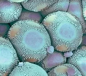
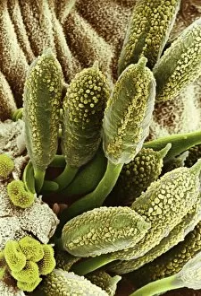
Rust fungus infection. Coloured scanning electron micrograph (SEM) of spore-bearing aecia from a rust (Puccinia lagenophora) fungus, on a common groundsel (Senecio vulgaris) plant

Background imageMycological Collection: Yeast cells, SEM

Yeast cells, SEM
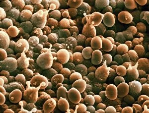
Yeast cells. Coloured scanning electron micrograph (SEM) of cells of bakers yeast (Saccharomyces cerevisiae) from part of a dried, commercial yeast pellet

Background imageMycological Collection: Mushroom anatomy, diagram

Mushroom anatomy, diagram
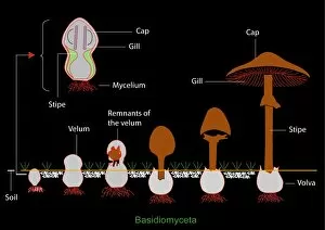
Mushroom anatomy. Diagram of the anatomy of Basidiomyceta mushrooms, and their growth from a fungal mycelium in the soil (left) to a full-grown mushroom (right)

Background imageMycological Collection: Mouldy lemon

Mouldy lemon. Penicillium sp. fungus growing on a lemon (Citrus limon). The fungus has spread over the surface of the fruit, producing a network (mycelium, white) of fine filaments (hyphae)

Background imageMycological Collection: Freckled dapperling (Lepiota aspera) C018 / 1771

Freckled dapperling (Lepiota aspera) C018 / 1771
Freckled dapperling (Lepiota aspera) in a woodland clearing

Background imageMycological Collection: Rust fungus on a rose leaf, SEM C017 / 7132

Rust fungus on a rose leaf, SEM C017 / 7132
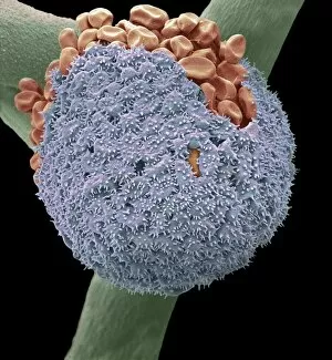
Rust fungus on a rose leaf. Coloured scanning electron micrograph (SEM) of rust fungus (Phragmidium sp.) spores emerging from a rose (Rosa sp.) leaf (brown)

Background imageMycological Collection: Type I topoisomerase protein bound to DNA F007 / 9893

Type I topoisomerase protein bound to DNA F007 / 9893
Type I topoisomerase bound to DNA. Molecular model showing a type I topoisomerase molecule (blue) bound to a strand of DNA (deoxyribonucleic acid, yellow and red)

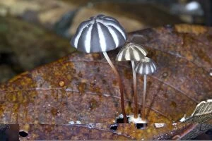
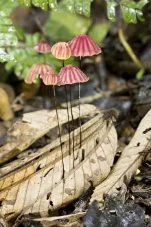
Background imageMycological Collection: Tropical mushrooms C014 / 0952

Tropical mushrooms C014 / 0952
Tropical mushrooms growing in leaf litter. Photographed in Rio Bigal Biological Reserve, Amazon Rainforest, Ecuador

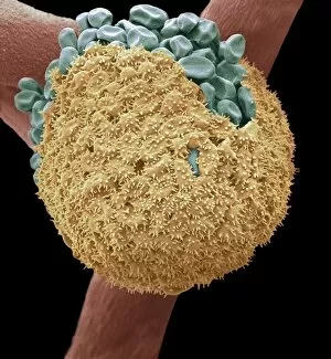
Background imageMycological Collection: Fungal spores on pollen grain, SEM

Fungal spores on pollen grain, SEM
Fungal spores on pollen grain. Coloured scanning electron micrograph (SEM) showing fungal spores (round) on a pollen grain from a goji berry (wolfberry, Lycium sp.) plant

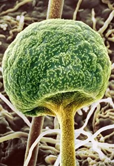
Background imageMycological Collection: Bread mould, SEM C017 / 7127

Bread mould, SEM C017 / 7127
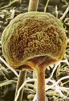
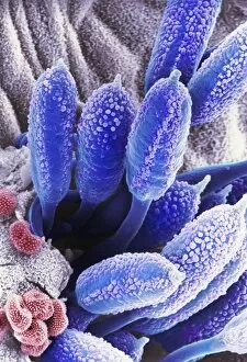
Bread mould. Coloured scanning electron micrograph (SEM) of a sporangium of a bread mould (Mucor sp.). Sporangia are asexual reproductive structures where the moulds spores develop

Background imageMycological Collection: Bread mould, SEM C017 / 7129

Bread mould, SEM C017 / 7129
Bread mould. Coloured scanning electron micrograph (SEM) of a sporangium of a bread mould (Mucor sp.). Sporangia are asexual reproductive structures where the moulds spores develop

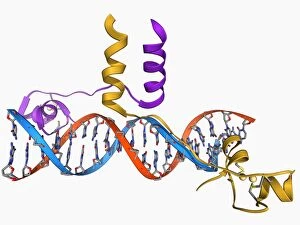
Background imageMycological Collection: Yeast DNA recognition, molecular model F006 / 9282

Yeast DNA recognition, molecular model F006 / 9282
Yeast DNA recognition. Computer model showing a GAL4 transcription activator protein bound to a yeast DNA (deoxyribonucleic acid) molecule (red and blue)

Background imageMycological Collection: Cup lichen (Cladonia sp. )

Cup lichen (Cladonia sp. )
Cup lichen (Cladonia sp.) growing on a stone. Lichens are symbiotic organisms formed of a fungus and an alga that mutually benefit one another. Photographed in the UK

Background imageMycological Collection: Pine cone on lichen

Pine cone on lichen. Close-up of a cone from a pine (Pinus sp.) tree on reindeer lichen (Cladonia portentosa). Photographed in the UK

Background imageMycological Collection: Black chanterelle mushrooms

Black chanterelle mushrooms (Craterellus cornucopioides). Photographed in the New Forest, Hampshire, UK

Background imageMycological Collection: Bread mould, SEM C016 / 9051

Bread mould, SEM C016 / 9051
Bread mould. Coloured scanning electron micrograph (SEM) of a black bread mould (Rhizopus stolonifer) sporangium (orange, spherical)

Background imageMycological Collection: Curvularia geniculata fungus, SEM C016 / 9389

Curvularia geniculata fungus, SEM C016 / 9389
Curvularia geniculata fungus. Coloured scanning electron micrograph (SEM) of a colony of the filamentous fungus Curvularia geniculata. This hyphomycete fungus is a pathogen of many plant species

Background imageMycological Collection: Apple tree fungus, SEM C016 / 9416

Apple tree fungus, SEM C016 / 9416
Apple tree fungus. Coloured scanning electron micrograph (SEM) of fungal spores bursting through a lesion on the surface of a leaf from an apple (Malus domestica) tree

Background imageMycological Collection: Mould spores, SEM C016 / 9419

Mould spores, SEM C016 / 9419
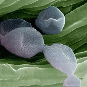
Mould spores. Coloured scanning electron micrograph (SEM) of mould (fungal) spores (blue) on food (green)

Background imageMycological Collection: Bread mould, SEM C016 / 9052

Bread mould, SEM C016 / 9052
Bread mould. Coloured scanning electron micrograph (SEM) of a black bread mould (Rhizopus stolonifer) sporangium (blue, spherical)

Background imageMycological Collection: Mushrooms spores, SEM C016 / 9450

Mushrooms spores, SEM C016 / 9450
Mushrooms spores. Coloured scanning electron micrograph (SEM) of budding spores in the basidium of an Agaricus bisporus fungus

Background imageMycological Collection: Fungus on orchid C016 / 7748

Fungus on orchid C016 / 7748
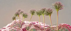
Fungus on orchid. Close-up of a tropical fungus growing on the leaves of an orchid (family Orchidaceae). Photographed in Yasuni National Park, Amazon rainforest, Ecuador

Background imageMycological Collection: Mould spores, SEM C016 / 9418

Mould spores, SEM C016 / 9418
Mould spores. Coloured scanning electron micrograph (SEM) of mould (fungal) spores (blue) on food (green)

Background imageMycological Collection: Thrush infection of the tongue, SEM C016 / 9091

Thrush infection of the tongue, SEM C016 / 9091
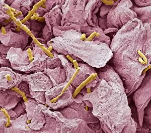
Thrush infection of the tongue. Coloured scanning electron micrograph (SEM) of the surface of a tongue infected with Candida sp. fungus (yellow), known as yeast infection, thrush, or candidiasis

Background imageMycological Collection: Apple tree fungus, SEM C016 / 9417

Apple tree fungus, SEM C016 / 9417
Apple tree fungus. Coloured scanning electron micrograph (SEM) of fungal spores bursting through a lesion on the surface of a leaf from an apple (Malus domestica) tree

Background imageMycological Collection: Bread mould, SEM C016 / 9050

Bread mould, SEM C016 / 9050
Bread mould. Coloured scanning electron micrograph (SEM) of a black bread mould (Rhizopus stolonifer) sporangium (green, spherical)

Background imageMycological Collection: Tropical mushrooms (Marasmius sp. ) C016 / 6240

Tropical mushrooms (Marasmius sp. ) C016 / 6240
Tropical mushrooms. Close-up of an agaric (Marasmius sp.) fungus growing in the leaf litter of the forest floor. Photographed in Yasuni National Park, Amazon rainforest, Ecuador

Background imageMycological Collection: Tropical mushrooms C016 / 6248

Tropical mushrooms C016 / 6248
Tropical mushrooms. Close-up of a fungus growing on a rotting log on the forest floor. Photographed in Yasuni National Park, Amazon rainforest, Ecuador

Background imageMycological Collection: Ant on a toadstool C016 / 6249

Ant on a toadstool C016 / 6249
Ant on a toadstool. Bulldog ant (Myrmecia sp.) on a Marasimus sp. toadstool on the rainforest floor. Photographed in Yasuni National Park, Amazon rainforest, Ecuador

Background imageMycological Collection: Bioluminescent mushrooms C016 / 6238

Bioluminescent mushrooms C016 / 6238
Bioluminescent mushrooms. Close-up of the bioluminescing fruiting bodies (mushrooms) of a tropical fungus growing on a rotting branch on the floor of a rainforest

Background imageMycological Collection: Fungus (Lycoperdon coliforme) C016 / 5988

Fungus (Lycoperdon coliforme) C016 / 5988
Fungus (Lycoperdon coliforme). Plate 1 from Drawings of Submerged Algae (1800) by Mary Dawson Turner. Held in the Botany Library at the Natural History Museum, London, UK

Background imageMycological Collection: Mushroom, SEM C015 / 9162

Mushroom, SEM C015 / 9162
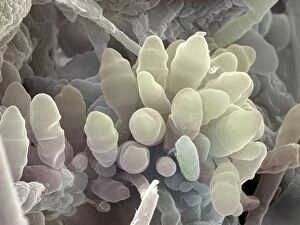
Mushroom, coloured scanning electron micrograph (SEM). Gills are seen on the underside of the mushrooms cap. These are the structures from which reproductive spores are shed

Background imageMycological Collection: Mushroom, SEM C015 / 9161

Mushroom, SEM C015 / 9161
Mushroom, coloured scanning electron micrograph (SEM). Gills are seen on the underside of the mushrooms cap. These are the structures from which reproductive spores are shed

Background imageMycological Collection: Rust fungus on a rose leaf, SEM C017 / 7131

Rust fungus on a rose leaf, SEM C017 / 7131
Rust fungus on a rose leaf. Coloured scanning electron micrograph (SEM) of rust fungus (Phragmidium sp.) spores emerging from a rose (Rosa sp.) leaf (brown)

Background imageMycological Collection: Bread mould, SEM C017 / 7128

Bread mould, SEM C017 / 7128
Bread mould. Coloured scanning electron micrograph (SEM) of a sporangium of a bread mould (Mucor sp.). Sporangia are asexual reproductive structures where the moulds spores develop

Background imageMycological Collection: Rust fungus on a rose leaf, SEM C017 / 7130

Rust fungus on a rose leaf, SEM C017 / 7130
Rust fungus on a rose leaf. Coloured scanning electron micrograph (SEM) of rust fungus (Phragmidium sp.) spores emerging from a rose (Rosa sp.) leaf (mauve)

Background imageMycological Collection: Type I topoisomerase bound to DNA C014 / 0862

Type I topoisomerase bound to DNA C014 / 0862
Type I topoisomerase bound to DNA. Molecular model showing a type I topoisomerase molecule (khaki) bound to a strand of DNA (deoxyribonucleic acid, pink and green)

Background imageMycological Collection: Type I topoisomerase bound to DNA C014 / 0883

Type I topoisomerase bound to DNA C014 / 0883
Type I topoisomerase bound to DNA. Molecular model showing a type I topoisomerase molecule (green) bound to a strand of DNA (deoxyribonucleic acid, pink and blue)

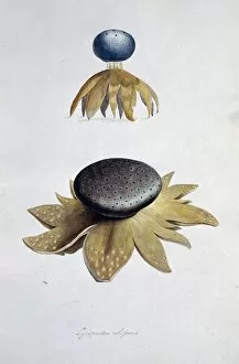
Background imageMycological Collection: Earthstar (Geastrum sp. ) fungus C014 / 0950

Earthstar (Geastrum sp. ) fungus C014 / 0950
Earthstar (Geastrum sp.) fungus in leaf litter. This fungus releases its spores from the central spherical structure. Photographed in Rio Bigal Biological Reserve, Amazon Rainforest, Ecuador

Background imageMycological Collection: Parasitic fungus on a weevil C014 / 0951

Parasitic fungus on a weevil C014 / 0951
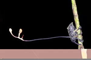
Parasitic fungus on a weevil. Fruiting body of a Cordyceps sp. fungus (left) growing out of the body of a weevil (superfamily Curculionoidea, right) clinging to a plant stem


All Professionally Made to Order for Quick Shipping

"Mycological Marvels: Exploring the Fascinating World of Fungi" Step into a realm where bread mould becomes a captivating subject under the scanning electron microscope (SEM). Witness the intricate details and mesmerizing patterns that adorn these seemingly ordinary fungi. Transport yourself to the early 1900s, as you gaze upon a botanical plate depicting both Good and Bad Mushrooms. Delight in the vibrant colors captured through lithography, showcasing nature's diversity in mushroom species. Discover Hyprophyllum aquifolii, elegantly displayed on Plate 38 from Iconographie des Champignons de J. J. , its delicate form beckoning you closer. Admire Hypodendrums fagi and queris on Plate 138, their unique features highlighted with precision by J. J. 's artistic hand. Marvel at Hypophyllum campestre or the field mushroom on Plate 130 from 'Iconographie des Champignons de J. J. , ' an exquisite representation of this culinary delight found in meadows. Let your imagination run wild as you envision Tubiporus cepa on Plate 176, enticingly illustrated for Traite des Champignons', promising a feast for those who dare to indulge. Delve deeper into Tubiporus esculentus on Plate 168 from Iconographie des Champignons de J. J. , marveling at its edible allure and contemplating its potential uses in gastronomy. Witness fungal fruiting bodies come alive before your eyes through stunning illustrations that capture their beauty and complexity. Explore another Botanical plate depicting Good and Bad Mushrooms from c. 1900 - a testament to mankind's fascination with these enigmatic organisms throughout history. Uncover Rust fungus infection through SEM imagery, revealing both the destructive power and awe-inspiring intricacy of nature's battles within microscopic realms. And finally, ponder upon a mouldy lemon.