
Muscle Contraction Collection
"Exploring the Intricate Dance of Muscle Contraction: A Captivating Artwork" This captivating artwork delves into the fascinating world of muscle contraction


All Professionally Made to Order for Quick Shipping
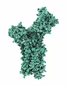
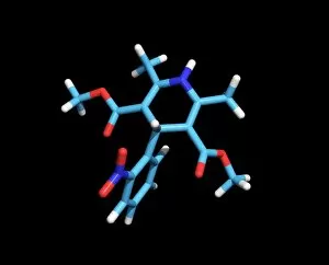
"Exploring the Intricate Dance of Muscle Contraction: A Captivating Artwork" This captivating artwork delves into the fascinating world of muscle contraction, showcasing its complexity and beauty. From a satirical depiction of foot cramps to intricate molecular structures, this piece encapsulates the essence of this physiological phenomenon. At its core, muscle contraction involves various key players. The Glycogen phosphorylase molecule (F006/9775) and Molecular motor protein (F006/9618) work in tandem to initiate and regulate the process. Meanwhile, Calcium-binding protein molecule (F006/9567) plays a crucial role in transmitting signals that trigger contractions. The artwork also highlights the importance of calcium regulation through proteins like Molecular motor protein (F006/9537), Calcium ATPase ion pump (F006/9507), and Calcium pumping ATPase muscle enzyme (F006/9377). These molecules ensure precise control over calcium levels within muscles, allowing for efficient contractions. Additionally, Myosin molecule (F006/9255) takes center stage as it interacts with actin filaments during muscle contraction. Its counterpart, Myosin fragment molecule (F006/9245), showcases a closer look at this essential component's structure and function. Drawing inspiration from Duchenne's physiognomy studies in the 1860s, this artwork pays homage to early scientific investigations into muscular physiology. It serves as a reminder of how far we have come in understanding these intricate processes that enable our bodies to move. Through vibrant colors and meticulous details, this artwork invites viewers on an immersive journey into the world of muscle contraction. It sparks curiosity about our own bodies' capabilities while celebrating the artistry found within science itself.