
Mouth Parts Collection
"Mouth Parts: A Fascinating World Revealed Through Microscopic Illustrations" Step into the intricate world with these captivating illustrations


All Professionally Made to Order for Quick Shipping
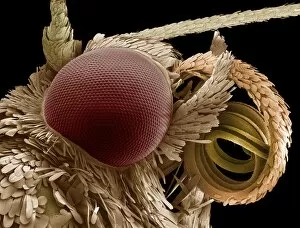
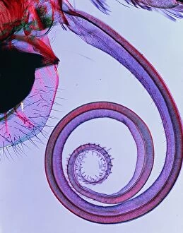
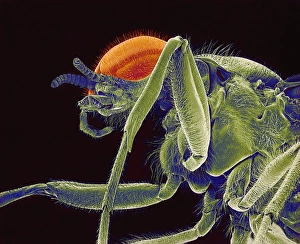
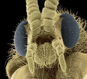
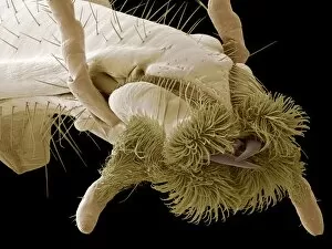
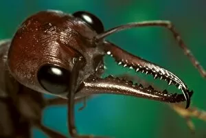
"Mouth Parts: A Fascinating World Revealed Through Microscopic Illustrations" Step into the intricate world with these captivating illustrations. Dating back to the 18th century, these hand-coloured engravings and microscopically detailed artworks offer a glimpse into the diverse anatomy of various creatures. In an enchanting illustration from 1790, we witness the delicate intricacies of a spider's mouth parts. Each tiny component is meticulously depicted, showcasing nature's remarkable design. Similarly, another engraving from the same era reveals multiple spiders in all their glory, highlighting their unique adaptations for feeding. Moving on to smaller organisms, an illustration from Musement Microscopique (1764) showcases an aphid's mouth parts. This microscopic marvel demonstrates how even seemingly insignificant creatures possess extraordinary structures that enable them to thrive. Garden enthusiasts will find interest in yet another illustration from Musement Microscopique which portrays various garden pests and their distinctive mouth parts. These depictions serve as a reminder that understanding such details can aid in pest control strategies while appreciating nature's diversity. Zooming further into this hidden realm, scanning electron microscope (SEM) images provide astonishingly detailed views of specific species' mouth parts. From the sheep tick to the black vine weevil and dog flea – each image captures minute features that contribute to their survival and interaction with hosts. Not limited to arthropods alone, our exploration extends towards moths and butterflies too. SEM imagery unveils mesmerizing close-ups of moth eyes coupled with proboscises used for feeding purposes. Delicate patterns adorn these organs while demonstrating nature's ingenuity at work. Lastly, we delve into butterfly anatomy through polarized light microscopy (PLM). The sucking proboscis of a butterfly named P. brassicae takes center stage here – its structure revealed in stunning detail under specialized lighting techniques. These captivating illustrations remind us that there is much more to creatures' mouth parts than meets the eye.