Fraud Blocker Skip to main content

Microscopic Collection (page 35)

"Exploring the Microscopic World: A Glimpse into Nature's Hidden Wonders" Step into a realm unseen by the naked eye

Background imageMicroscopic Collection: Scanning Electron Micrograph (SEM): Spectacle Moth, Magnification x 200 (A4 size: 29. 7 cm width)

Scanning Electron Micrograph (SEM): Spectacle Moth, Magnification x 200 (A4 size: 29. 7 cm width)
LRDS-113 Spectacle Moth - caterpillars hatching Scanning Electron Micrograph (SEM) Abrostola tripartita Magnification x 200 (A4 size: 29.7 cm width) Coloured by hand to enhance natural features

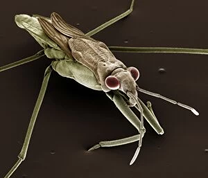
Background imageMicroscopic Collection: Scanning Electron Micrograph (SEM): Pond Skater, Magnification x 40 (A4 size: 29. 7 cm width)

Scanning Electron Micrograph (SEM): Pond Skater, Magnification x 40 (A4 size: 29. 7 cm width)
LRDS-111 Pond Skater Scanning Electron Micrograph (SEM) Gerris lacustris Magnification x 40 (A4 size: 29.7 cm width) Coloured by hand to enhance natural features

Background imageMicroscopic Collection: Scanning Electron Micrograph (SEM): Psocid, Magnification x 100 (A4 size: 29. 7 cm width)

Scanning Electron Micrograph (SEM): Psocid, Magnification x 100 (A4 size: 29. 7 cm width)
LRDS-104 Psocid Scanning Electron Micrograph (SEM) Lepinotus sp. Magnification x 100 (A4 size: 29.7 cm width) Coloured by hand to enhance natural features

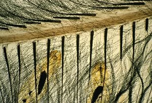
Background imageMicroscopic Collection: Feather Louse - on feather. Magnification x10

Feather Louse - on feather. Magnification x10
JLMO-2353 Feather Louse - on feather. Magnification x10 Nirmus merulensis John Mason Please note that prints are for personal display purposes only and may not be reproduced in any way

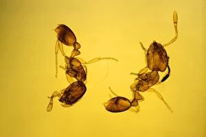
Background imageMicroscopic Collection: Pharaoh's Ants Magnification x10

Pharaoh's Ants Magnification x10
JLMO-2351 Pharaoh's Ants Magnification x10 Monomorium pharaonis John Mason Please note that prints are for personal display purposes only and may not be reproduced in any way

Background imageMicroscopic Collection: Anwyl Bromeliads - water is trapped under the scaly on the surface of the plant

Anwyl Bromeliads - water is trapped under the scaly on the surface of the plant
PPG-1651 Anwyl Bromeliads - water is trapped under the scaly on the surface of the plant Tillandia streptophylla Pascal Goetgheluck Please note that prints are for personal display purposes only

Background imageMicroscopic Collection: Bladderwort - Bladder-like sac, ready to suck a worm in. South Africa

Bladderwort - Bladder-like sac, ready to suck a worm in. South Africa
PPG-1650 Bladderwort - bladder-like sac, ready to suck a worm in South Africa Utricularia sandersonii Pascal Goetgheluck Please note that prints are for personal display purposes only

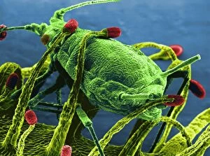
Background imageMicroscopic Collection: Drosera pulchella rosea'. Drosera. Greenfly caught by the tentacles

Drosera pulchella rosea'. Drosera. Greenfly caught by the tentacles. (Electronic Scanning Microscopy)
PPG-1644 Drosera pulchella rosea, Greenfly caught by the tentacles Drosera pulchella rosea Electronic Scanning Microscopy Pascal Goetgheluck Please note that prints are for personal display purposes

Background imageMicroscopic Collection: Sheep Ked / Tick

Sheep Ked / Tick
JC-600 INSECTS - Sheep Ked / Sheep Tick magnified x8 Melophagus ovina John Clegg Please note that prints are for personal display purposes only and may not be reproduced in any way

Background imageMicroscopic Collection: Bed Bug

Bed Bug
JC-598 Bed Bug - male; x 3 magnification Cimex lectularius John Clegg Please note that prints are for personal display purposes only and may not be reproduced in any way

Background imageMicroscopic Collection: A Drop of London Water

A Drop of London Water
THE WONDERS OF A LONDON WATER DROP A water drop as it would be seen under the Molecular Magnifier. It is home to many hideous microscopic creatures

Background imageMicroscopic Collection: Cholera Vibrio

Cholera Vibrio
Vibrio (or vibrion) of cholera, discovered by Koch, 1883

Background imageMicroscopic Collection: Microscopic Objects

Microscopic Objects
A variety of living and non- living objects magnified through a microscope


All Professionally Made to Order for Quick Shipping

"Exploring the Microscopic World: A Glimpse into Nature's Hidden Wonders" Step into a realm unseen by the naked eye, where tiny creatures and intricate structures come to life. In Picture No. 10851646, we are captivated by the Scanning Electron Micrograph (SEM) of a Praying Mantis, magnified x30. Its delicate features and mesmerizing details reveal a world within our reach. Moving on to another fascinating creature, the Tardigrade or 'Water Bear', magnified x1250 in an SEM image. This resilient micro-animal showcases its remarkable adaptability under extreme conditions. It reminds us that even in the tiniest forms of life, strength can be found. The Fruit Fly takes center stage next with an SEM image at x300 magnification. Despite its minuscule size, this common insect holds secrets waiting to be discovered under closer inspection. Its intricate body structure hints at its role in nature's grand tapestry. Diatoms from marine plankton samples grace our vision next - their elegant shapes and patterns resembling miniature works of art created by nature itself. These microscopic organisms play a vital role in Earth's ecosystems as primary producers. An illustration of a Flea C017/3435 introduces us to yet another hidden world beneath our feet - one filled with parasites and symbiotic relationships that shape entire ecosystems on both macro and micro scales. Diving deeper into marine diatoms at x25 magnification reveals their stunning beauty – intricate designs etched onto tiny shells floating through vast oceans, silently contributing to Earth's biodiversity. Anton van Leeuwenhoek's observations of Animalcules circa 1795 remind us of humanity's earliest encounters with microscopic lifeforms – pioneering discoveries that forever changed our understanding of biology and paved the way for modern scientific advancements. A Macro Photograph unveils Paenibacillus bacteria colonies thriving amidst laboratory-imposed stresses, showcasing their ability to adapt and survive in challenging environments.