Fraud Blocker Skip to main content

Microscope Collection (page 6)

"Unveiling the Unseen: Exploring the Microscopic World" Step into a realm of hidden wonders

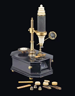
Background imageMicroscope Collection: Compound box-type microscope, c. 1775 (wood, metal & glass) (see also 1113960-1)

Compound box-type microscope, c. 1775 (wood, metal & glass) (see also 1113960-1)
1113959 Compound box-type microscope, c.1775 (wood, metal & glass) (see also 1113960-1) by Brander, Georg Friedrich (18th century); Private Collection; (add.info.: BOX: 11 1/8 X 8 X 4 1/2 IN)

Background imageMicroscope Collection: Microscope, c. 1690 (silver & glass) (see also 1121456-60)

Microscope, c. 1690 (silver & glass) (see also 1121456-60)
1121459 Microscope, c.1690 (silver & glass) (see also 1121456-60) by Leeuwenhoek, Antoni van (1632-1723); Private Collection; (add.info.: The lens held between two riveted silver plates)

Background imageMicroscope Collection: Fighting the Plague at Poona, the General Plague Hospital (b / w photo)

Fighting the Plague at Poona, the General Plague Hospital (b / w photo)
1039410 Fighting the Plague at Poona, the General Plague Hospital (b/w photo) by English photographer, (19th century) (after); Private Collection; (add.info.: Fighting the Plague at Poona)

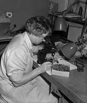
Background imageMicroscope Collection: The Entomologist

The Entomologist
Nick Walton

Background imageMicroscope Collection: Parthenogenesis

Parthenogenesis
Christophe Kiciak

Background imageMicroscope Collection: 1095333

1095333

Background imageMicroscope Collection: CoronaVirus

CoronaVirus
Dmitry Skvortsov

Background imageMicroscope Collection: Neolucanus delicatus

Neolucanus delicatus
riyad_hamzi

Background imageMicroscope Collection: Esterbrook Pen Factory 21st November 1967 Pen nibs being modified following quality

Esterbrook Pen Factory 21st November 1967 Pen nibs being modified following quality control checks at the Esterbrook Pen factory at Moland Street, Birmingham

Background imageMicroscope Collection: Schoolboys will soon be getting to know George, the skeleton, in biology lessons

Schoolboys will soon be getting to know George, the skeleton, in biology lessons, but teacher may not allow him to smoke when working with a microscope

Background imageMicroscope Collection: Professor Andrew Fielding Huxley of the University College who shares the Nobel Prize

Professor Andrew Fielding Huxley of the University College who shares the Nobel Prize. October 1963 P005291

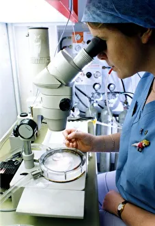
Background imageMicroscope Collection: Senior embryologist Jane Skelton using state-of-the-art micro manipulation equipment at

Senior embryologist Jane Skelton using state-of-the-art micro manipulation equipment at Walsgrave hospitals assisted conception unit, Coventry. 7th January 1994

Background imageMicroscope Collection: A contribution by the Manchester Microscopical Society were these two microscopes

A contribution by the Manchester Microscopical Society were these two microscopes. On left is a Ross microscope of approx 1850; the unit in the centre being the paraffin lamp used for illumination of

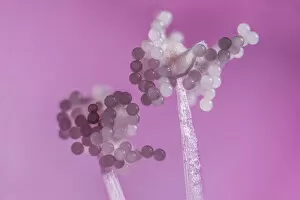
Background imageMicroscope Collection: Hollyhock (Alcea rosea), pollen grains on anthers, aprox 12x magnification, Monmouthshire

Hollyhock (Alcea rosea), pollen grains on anthers, aprox 12x magnification, Monmouthshire, Wales, UK

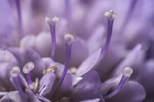
Background imageMicroscope Collection: Small scabious (Scabiosa columbaria) with three tiny (1mm) Thrips (Order Thysanoptera

Small scabious (Scabiosa columbaria) with three tiny (1mm) Thrips (Order Thysanoptera) on a petal, Monmouthshire, Wales, UK

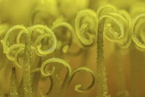
Background imageMicroscope Collection: Dandelion (Taraxacum officinale) stigma open to receive pollen for fertilization

Dandelion (Taraxacum officinale) stigma open to receive pollen for fertilization, Monmouthshire, Wales, UK, July

Background imageMicroscope Collection: Thereza, 1853-56. Creator: John Dillwyn Llewelyn

Thereza, 1853-56. Creator: John Dillwyn Llewelyn
Thereza, 1853-56

Background imageMicroscope Collection: Nursing classroom

Nursing classroom
An empty classroom at the City & District Infirmary York probationer school founded in 1923. Table at centre with small microscope, and bed on left hand side with large doll. Date: circa 1923

Background imageMicroscope Collection: Scientist Loves Cadburys

Scientist Loves Cadburys
An archetypal scientist, with a flowing beard and surrounded by apparatus, studies a sample of Cadburys Cocoa; he of course concludes that it is the finest available. Date: circa 1890

Background imageMicroscope Collection: WINKELS MICROSCOPE

WINKELS MICROSCOPE
Winkels microscope Date: late 19th century

Background imageMicroscope Collection: MICROSCOPE LATE C19

MICROSCOPE LATE C19
Date: late 19th century

Background imageMicroscope Collection: VARIOUS MICROSCOPES

VARIOUS MICROSCOPES
A selection of microscopes Date: 1797

Background imageMicroscope Collection: MICROSCOPE / ENC. BRIT

MICROSCOPE / ENC. BRIT
A selection of microscopes

Background imageMicroscope Collection: ROBERT KOCH / IN KIMBERLEY

ROBERT KOCH / IN KIMBERLEY
HEINRICH HERMANN ROBERT KOCH German physician and pioneer bacteriologist in search of the Rinderpest microbe at Kimberley Date: 1843 - 1910

Background imageMicroscope Collection: Louis Pasteur in his laboratory

Louis Pasteur in his laboratory
Louis Pasteur (1822-1895), French chemist and microbiologist, in his laboratory. Date: 1885

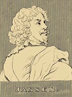
Background imageMicroscope Collection: Jansen, (1585-c1632), 1830. Creator: Unknown

Jansen, (1585-c1632), 1830. Creator: Unknown
Jansen, (1585-c1632), 1830. Zacharias Janssen (1585-c1632) Dutch spectacle-maker associated with invention of the first optical telescope

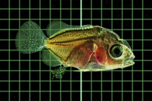
Background imageMicroscope Collection: Picture No. 12479490

Picture No. 12479490
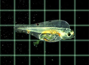
Marine fish larvae eat microplastics. Small pieces Date:

Background imageMicroscope Collection: Picture No. 12479491

Picture No. 12479491
Marine fish larvae eat microplastics. Small pieces Date:

Background imageMicroscope Collection: Dr. Atkinson in his Laboratory, 15 September 1911, (1913). Artist: Herbert Ponting

Dr. Atkinson in his Laboratory, 15 September 1911, (1913). Artist: Herbert Ponting
Dr. Atkinson in his Laboratory, 15 September 1911, (1913). Surgeon and parasitologist Edward Atkinson (1881-1929) led the party which found the tent with the bodies of Scott, Bowers and Wilson

Background imageMicroscope Collection: Debenham, Gran, and Taylor in their Cubicle, 18 May 1911, (1913). Artist: Herbert Ponting

Debenham, Gran, and Taylor in their Cubicle, 18 May 1911, (1913). Artist: Herbert Ponting
Debenham, Gran, and Taylor in their Cubicle, 18 May 1911, (1913). Geologists Frank Debenham (1883-1965) and T Griffith Taylor (1880-1963), and ski expert Tryggve Gran (1888-1980)

Background imageMicroscope Collection: The Popular Science Educator front cover, 1935

The Popular Science Educator front cover, 1935
The Popular Science Educator front cover, . From The Popular Science Educator, Volume 1, edited by Charles Ray. [The Amalgamated Press, Ltd. London, ]

Background imageMicroscope Collection: Santiago Ramon y Cajal (1852-1934), Spanish physician and researcher, Nobel

Santiago Ramon y Cajal (1852-1934), Spanish physician and researcher, Nobel Prize in Medicine in 1906

Background imageMicroscope Collection: Electronic microscope of Siemens transmission ST100F

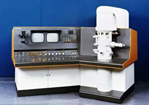
Electronic microscope of Siemens transmission ST100F

Background imageMicroscope Collection: A lab tachnician with a Reichter Microscope at a steelworks, Sheffield, South Yorkshire, 1962

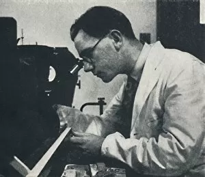
A lab tachnician with a Reichter Microscope at a steelworks, Sheffield, South Yorkshire, 1962. A lab tachnician checks the quality of tungsten carbide with at the Edgar Allen Steel Co

Background imageMicroscope Collection: Research worker with microscope (testing the structure of an aluminium alloy), 1941

Research worker with microscope (testing the structure of an aluminium alloy), 1941. Artist: Cecil Beaton
Research worker with microscope (testing the structure of an aluminium alloy), 1941. From Air of Glory, by Cecil Beaton. [His Majestys Stationery Office, London, 1941]

Background imageMicroscope Collection: Apprentice with microscope (testing the hardness of pistons), 1941. Artist: Cecil Beaton

Apprentice with microscope (testing the hardness of pistons), 1941. Artist: Cecil Beaton
Apprentice with microscope (testing the hardness of pistons), 1941. From Air of Glory, by Cecil Beaton. [His Majestys Stationery Office, London, 1941]

Background imageMicroscope Collection: Microscope, 1912

Microscope, 1912. From The Book of Sports & Pastimes for Young People by J. K. Benson. [C. Arthur Pearson, Ltd, London, 1912]

Background imageMicroscope Collection: Emblem of the Paris International Exhibition, 1867

Emblem of the Paris International Exhibition, 1867. A print from The Illustrated London News, (20 July 1867)

Background imageMicroscope Collection: Ilya Ilyich Mechnikov, Russian microbiologist, c1900s(?). Artist: Nadar

Ilya Ilyich Mechnikov, Russian microbiologist, c1900s(?). Artist: Nadar
Ilya Ilyich Mechnikov, Russian microbiologist, c1900s(?). Mechnikov (1845-1916) continued the work of Louis Pasteur, specialising in the study of the immune system

Background imageMicroscope Collection: Mr Frank Crisp, 1890. Artist: Spy

Mr Frank Crisp, 1890. Artist: Spy
Mr Frank Crisp, 1890. Crisp (c1853-1919), an English Limited Liability Lawyer one of whose personal interests was microscopy, acted as Secretary of the Royal Microscopical Society

Background imageMicroscope Collection: Frontispiece of Ontledigen en Ondekkigen... Brieven by Anton van Leeuwenhoek, 1686

Frontispiece of Ontledigen en Ondekkigen... Brieven by Anton van Leeuwenhoek, 1686
Frontispiece of Ontledigen en Ondekkigen...Brieven by Dutch microscopist Anton van Leeuwenhoek, 1686. Leeuwenhoek (1632-1723)

Background imageMicroscope Collection: Dorothea Klumpke Roberts, American mathematician and astronomer, 1903

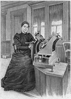
Dorothea Klumpke Roberts, American mathematician and astronomer, 1903. Roberts (1861-1942) at work on the Carte du Ciel at the Paris Observatory

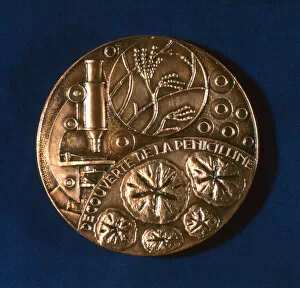
Background imageMicroscope Collection: Medal commemorating the discovery of penicillin, 1945

Medal commemorating the discovery of penicillin, 1945
Medal commemorating the discovery of penicillin. Scottish bacteriologist Alexander Fleming (1881-1955) discovered penicillin in 1928

Background imageMicroscope Collection: Microscopes and microscopical objects, 1750

Microscopes and microscopical objects, 1750. I: Wilsons pocket microscope. II: Scroll microscope. III: Tripod microscope - improved form of Marshalls double microscope

Background imageMicroscope Collection: Hookes microscope with condenser for concentrating light, 1665

Hookes microscope with condenser for concentrating light, 1665. From left to right above are his barometer, refractometer for measuring refractive power of liquids, and lens-grinding machine


All Professionally Made to Order for Quick Shipping

"Unveiling the Unseen: Exploring the Microscopic World" Step into a realm of hidden wonders, where scientists like Rosalind Franklin and Santiago Ramon y Cajal have unraveled the mysteries through their microscopic lens. From Liver Fluke to Norovirus particles, these pioneers have paved the way for groundbreaking discoveries. Witness the intricate beauty of Snail teeth and Fat cells as they come alive under the microscope's gaze. Marvel at Lubbock's Graphic 84, a visual masterpiece capturing the essence of this microcosmic universe. Delve deeper into this microscopic world and encounter Plasmodium sp. , a malarial parasite that has plagued humanity for centuries. Explore E. Coli bacterium in all its glory, revealing both its potential dangers and scientific significance. Through microscope slide preparation techniques, we gain insight into complex organisms like never before. Discover how Fleming used his photographic skills to document crucial moments in microbiology history. The microscope acts as our window into an unseen dimension - it allows us to explore realms beyond our naked eye's reach, and is through this powerful tool that we continue to unravel nature's secrets one magnified image at a time. So join us on this journey "Under the Microscope, " where every slide holds a story waiting to be told – stories that shape our understanding of life itself.