
Microscope Slide Collection
"Exploring the Intricacies of the Human Brain: A Glimpse through Microscope Slides" Delving into the depths of scientific discovery


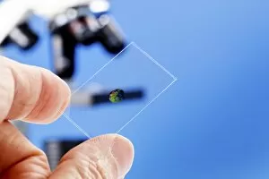


All Professionally Made to Order for Quick Shipping
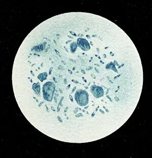
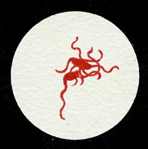
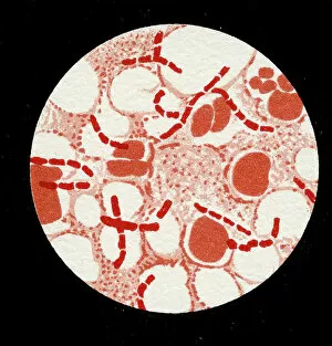
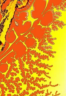
"Exploring the Intricacies of the Human Brain: A Glimpse through Microscope Slides" Delving into the depths of scientific discovery, microscope slides offer a mesmerizing view into the intricate world of microorganisms. These snapshots from 1906 provide a fascinating glimpse into various colonies and bacteria that have shaped our understanding of infectious diseases. One slide reveals a colony of Haemophilus influenzae, showcasing its unique structure and composition under magnification. Another showcases Mycobacterium leprae, offering insights into this ancient bacterium responsible for leprosy. The litho captures Micrococcus Gonorrhoea in all its glory, shedding light on one of humanity's most prevalent sexually transmitted infections. Streptococcus pneumoniae takes center stage in multiple slides - some with bubble capsules and others without - highlighting their distinct characteristics. Meanwhile, Spirochaetes Borrelia Recurrentis, causing Lyme disease, is captured within a blood sample; an eerie reminder of the havoc these bacteria can wreak on human health. Vibrio cholerae makes an appearance as well, reminding us of its role in devastating cholera outbreaks throughout history. Clostridium tetani showcases its spores which contribute to tetanus infections while Mycobacterium tuberculosis reminds us why it remains one of humanity's greatest challenges even today. Lastly, Staphylococcus pyogenes steals attention with captivating images capturing cell division and presenting colonies in their full splendor. Streptococcus pyogenes also graces another slide with its presence - a testament to how these microscopic organisms continue to shape our lives. These microscope slides serve as time capsules from over a century ago when scientists were just beginning to unravel the mysteries hidden within each tiny organism. They remind us that every breakthrough starts with curiosity and observation through lenses so small yet capable of revealing worlds beyond imagination.