Fraud Blocker Skip to main content

Microorganism Collection (page 5)

Microorganisms, the tiny life forms that exist all around us, are truly fascinating

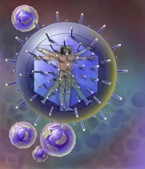
Background imageMicroorganism Collection: Vitruvian Man inside virus particle

Vitruvian Man inside virus particle

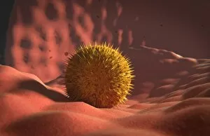
Background imageMicroorganism Collection: Microscopic view of Paramecium bursaria

Microscopic view of Paramecium bursaria, a species of ciliate protozoan that has a mutualistic symbiotic relationship with green algae called Zoochlorella

Background imageMicroorganism Collection: Microscopic view of protozoa

Microscopic view of protozoa. Leishmaniasis and dysentery are examples of diseases involving protozoa, often transmitted to humans by the bite of a parasite-carrying insect

Background imageMicroorganism Collection: Microscopic view of stem cell development

Microscopic view of stem cell development. In adult organisms, stem cells and progenitor cells act as a repair system for the body, replenishing adult tissues

Background imageMicroorganism Collection: Microscopic view of coronavirus

Microscopic view of coronavirus

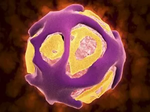
Background imageMicroorganism Collection: Conceptual image of cancer virus

Conceptual image of cancer virus

Background imageMicroorganism Collection: Microscopic view of Syphillis

Microscopic view of Syphillis. Syphilis is a sexually transmitted infection caused by the spirochete bacterium, Treponema pallidum

Background imageMicroorganism Collection: Microscopic view of athletes foot fungus

Microscopic view of athletes foot fungus. Athletes foot fungus, also known as ringworm of the foot and tinea pedis, is a fungal infection of the skin

Background imageMicroorganism Collection: Colorized scanning electron micrograph of a T lymphocyte

Colorized scanning electron micrograph of a T lymphocyte

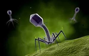
Background imageMicroorganism Collection: Microscopic view of bacteriophage

Microscopic view of bacteriophage

Background imageMicroorganism Collection: Structure of HIV

Structure of HIV

Background imageMicroorganism Collection: Grouping of virus particles

Grouping of virus particles

Background imageMicroorganism Collection: Microscopic view of Rickettsia

Microscopic view of Rickettsia. Rickettsia is a gram-negative bacteria carried by parasites such as ticks, fleas and lice

Background imageMicroorganism Collection: Microscopic view of corncob formation in dental plaque

Microscopic view of corncob formation in dental plaque, believed to be limited to strains of Corynebacterium matruchotii and Streptococcus sanguis

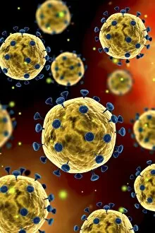
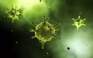
Background imageMicroorganism Collection: Conceptual image of the coronavirus

Conceptual image of the coronavirus. Coronaviruses primarily infect the upper respiratory and gastrointestinal tract

Background imageMicroorganism Collection: Microscopic view of virus

Microscopic view of virus

Background imageMicroorganism Collection: Microscopic view of herpes virus

Microscopic view of herpes virus

Background imageMicroorganism Collection: Conceptual image of influenza causing flu virus

Conceptual image of influenza causing flu virus

Background imageMicroorganism Collection: Close-up view of athletes foot fungus

Close-up view of athletes foot fungus, also known as ringworm of the foot and tinea pedis. Athletes foot fungus is a fungal infection of the skin

Background imageMicroorganism Collection: Group of Escherichia coli bacteria cells, known as E. Coli

Group of Escherichia coli bacteria cells, known as E. Coli
Group of Escherichia coli bacteria cells, commonly known as E. Coli. E. coli is a common type of bacteria that can get into food, like beef and vegetables

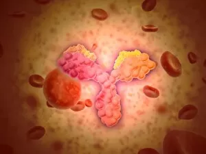
Background imageMicroorganism Collection: Microscopic view of human anitbodies with red blood cells

Microscopic view of human anitbodies with red blood cells. Human antibodies are the Y-shaped proteins used by the immune system to defend against foreign objects like bacteria and viruses

Background imageMicroorganism Collection: Virus landing on cell membrane

Virus landing on cell membrane

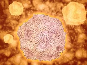
Background imageMicroorganism Collection: Microscopic view of rotavirus

Microscopic view of rotavirus. Rotavirus is the most common cause of severe diarrhea among infants and young children. It is a genus of double-stranded RNA virus in the family Reoviridae

Background imageMicroorganism Collection: Microscopic view of cell and virus

Microscopic view of cell and virus

Background imageMicroorganism Collection: Microscopic view of cancer virus

Microscopic view of cancer virus

Background imageMicroorganism Collection: Microscopic view of a group of Borrelia burgdorferi

Microscopic view of a group of Borrelia burgdorferi, the bacterial agent of Lyme disease transmitted by ticks

Background imageMicroorganism Collection: Microscopic view of Rubella virus

Microscopic view of Rubella virus
Microscopic view of Rubella. Rubella is an acute, contagious viral infection. While the illness is generally mild in children, it has serious consequences in pregnant women causing fetal death

Background imageMicroorganism Collection: Microscopic view of Legionella pneumophila

Microscopic view of Legionella pneumophila. Legionella pneumophila is a thin, aerobic, pleomorphic, flagellated, non-spore forming, gram-negative bacterium of the genus Legionella

Background imageMicroorganism Collection: Conceptual image of bacteria

Conceptual image of bacteria

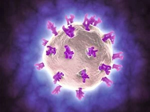
Background imageMicroorganism Collection: Conceptual image of common virus

Conceptual image of common virus

Background imageMicroorganism Collection: Conceptual image illustrating the process of bone metastasis

Conceptual image illustrating the process of bone metastasis

Background imageMicroorganism Collection: Microscopic view of Penicillium expansum

Microscopic view of Penicillium expansum, a mold fungi that effects plants such as apples and similar fruit. This fungus is known as blue mold

Background imageMicroorganism Collection: Conceptual image of the dengue virus

Conceptual image of the dengue virus. The dengue virus is a mosquito-borne RNA virus that causes dengue fever

Background imageMicroorganism Collection: Microscopic view of Yersinia

Microscopic view of Yersinia. Yersinia is a gram-negative, rod-shaped bacteria in the family Enterobacteriaceae

Background imageMicroorganism Collection: Conceptual image of salmonella

Conceptual image of salmonella. Salmonella is a genus of rod-shaped, gram-negative bacteria. There are two main species of Salmonella, Salmonella bongori and Salmonella enterica

Background imageMicroorganism Collection: Microscopic view of a microbe

Microscopic view of a microbe. Microbes are single-cell organisms so tiny that millions can fit into the eye of a needle

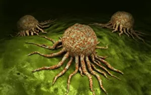
Background imageMicroorganism Collection: Microscopic view of tumor

Microscopic view of tumor

Background imageMicroorganism Collection: Conceptual image of flagellate bacterium

Conceptual image of flagellate bacterium

Background imageMicroorganism Collection: Microscopic view of bacteria

Microscopic view of bacteria

Background imageMicroorganism Collection: Malria spores in the human blood stream

Malria spores in the human blood stream
The malaria spores move from the mosquito to the human blood stream. the spores divide many times and produce large numbers of spores

Background imageMicroorganism Collection: Conceptual image of lyssavirus

Conceptual image of lyssavirus. Lyssavirus is a genus of viruses belonging to the family Rhabdoviridae. This group of RNA viruses includes the rabies virus traditionally associated with the disease

Background imageMicroorganism Collection: Conceptual image of a ubiquitous virus

Conceptual image of a ubiquitous virus. A ubiquitous virus is contagious in early childhood through the respiratory tract

Background imageMicroorganism Collection: Microscopic view of Henipavirus

Microscopic view of Henipavirus. Henipavirus is a established group of paramyxoviruses comprising the Hendra virus, Nipah virus and Cedar virus

Background imageMicroorganism Collection: Conceptual image of vibrio cholerae causing cholera

Conceptual image of vibrio cholerae causing cholera

Background imageMicroorganism Collection: Microscopic view of Canine Parvovirus

Microscopic view of Canine Parvovirus. Canine Parvovirus is a highly contagious viral disease that is one of the most common causes of diarrhea in dogs under 6 months of age

Background imageMicroorganism Collection: Influenza virus, glassy look

Influenza virus, glassy look

Background imageMicroorganism Collection: Conceptual image of common bacteria

Conceptual image of common bacteria

Background imageMicroorganism Collection: Paramecium -Paramecium-, dyed in different colours, permanent preparation, photomicrography

Paramecium -Paramecium-, dyed in different colours, permanent preparation, photomicrography


All Professionally Made to Order for Quick Shipping

Microorganisms, the tiny life forms that exist all around us, are truly fascinating. From HeLa cells to water bears and viruses like MRSA and RSV, these microscopic entities play a significant role in our lives. In the world of science, HeLa cells have been instrumental in numerous breakthroughs. Captured under a light micrograph (C017 / 8299), their intricate structures reveal the complexity of human biology. Another captivating image shows neutrophils engulfing MRSA bacteria, as seen through a scanning electron microscope (C018 / 8596). This powerful defense mechanism showcases how our immune system fights against harmful invaders. Water bears, also known as tardigrades, are remarkable creatures that can survive extreme conditions. A light micrograph (C016 / 8581) captures their unique appearance with their stout bodies and claw-like limbs. Equally intriguing is another image taken using a scanning electron microscope (SEM C016 / 9084), which reveals even more details about these resilient organisms. Viruses such as respiratory syncytial virus and flu virus particles have caused widespread infections throughout history. While one can only be observed under a microscope (microscopic view of human respiratory syncytial virus), the other is depicted through artwork showcasing its structure (flu virus particle, artwork F008 / 3245). These images remind us of the importance of vaccinations in preventing viral outbreaks. The spread of infections by sneezing is illustrated through an artistic representation (artwork C013 / 5949). It serves as a reminder to practice good hygiene to protect ourselves and others from contagious diseases. Chlamydia may be small but has significant consequences for those affected by it. A microscopic view highlights its presence within host cells—a stark reminder of the need for awareness and prevention efforts against sexually transmitted infections. Looking back at historical lithographs from 1906 brings attention to colonies formed by Haemophilus influenzae and Mycobacterium leprae.