Fraud Blocker Skip to main content

Microbial Collection

In the 19th century, microbial discoveries revolutionized the field of microbiology

Background imageMicrobial Collection: Salmonella bacteria, artwork C013 / 8818

Salmonella bacteria, artwork C013 / 8818
Salmonella bacteria, computer artwork. Salmonella sp. bacteria are gram-negative rod-shaped bacteria that have flagella (hair-like structures) that they use for locomotion

Background imageMicrobial Collection: Microbiology caricature, 19th century

Microbiology caricature, 19th century
Microbiology caricature. The researcher is handling jars labelled as bacterial cultures, while the magnified view at left shows a caricatured appearance of microbes as seen under a microscope

Background imageMicrobial Collection: Vinegar production, 19th century

Vinegar production, 19th century cutaway artwork. This is a trickling method developed in 1823 by the German chemist Schutzenbach. The barrel is divided into three sections

Background imageMicrobial Collection: Louis Pasteur (1822-95) and bacterial diseases, from

Louis Pasteur (1822-95) and bacterial diseases, from
CHT236774 Louis Pasteur (1822-95) and bacterial diseases, from Les Sciences Physiques et Naturelles, Paris, 1908 (colour litho) by French School

Background imageMicrobial Collection: Louis Pasteur, from The Century Illustrated Monthly Magazine

Louis Pasteur, from The Century Illustrated Monthly Magazine
2619188 Louis Pasteur, from The Century Illustrated Monthly Magazine, published 1884 (wood engraving) by Velten, H. (fl.1850-84); Private Collection; (add.info.: Louis Pasteur, 1822 A- 1895)

Background imageMicrobial Collection: Botulism bacteria

Botulism bacteria. Light micrograph of botulism bacteria (Clostridium botulinum). These bacteria occur naturally in soil. They produce botulinum toxin

Background imageMicrobial Collection: Anthrax antibiotics research

Anthrax antibiotics research. Antibiotic drugs (discs) being tested on anthrax bacteria (Bacillus anthracis) colonies being cultured on an agar growth medium in a petri dish

Background imageMicrobial Collection: Infectious bronchitis virus (IBV), TEM

Infectious bronchitis virus (IBV), TEM
Infectious bronchitis virus (IBV) particles, coloured transmission electron micrograph (TEM). This virus is a member of the coronavirus family

Background imageMicrobial Collection: Mouldy bread

Mouldy bread. The white areas are fungal mycelia, masses of thread-like structures (hyphae) that absorb nutrients. The blue-green areas are the fungal spores (reproductive structures)

Background imageMicrobial Collection: Thermophile bacterial mats at hotspring, Grand Prismatic Spring, Midway Geyser Basin

Thermophile bacterial mats at hotspring, Grand Prismatic Spring, Midway Geyser Basin, Yellowstone N. P. Wyoming, U. S
Thermophile bacterial mats at hotspring, Grand Prismatic Spring, Midway Geyser Basin, Yellowstone N.P. Wyoming, U.S.A. September

Background imageMicrobial Collection: Thermophile bacterial mats and steam rising from hotspring, with tourists on boardwalk

Thermophile bacterial mats and steam rising from hotspring, with tourists on boardwalk, Grand Prismatic Spring, Midway Geyser Basin, Yellowstone N.P. Wyoming, U.S.A. September

Background imageMicrobial Collection: Joseph Lister, British surgeon

Joseph Lister, British surgeon
Joseph Lister (1827-1912), British surgeon. Lister qualified as a surgeon in London in 1852. At that time surgery was usually followed by infection and high mortality rates

Background imageMicrobial Collection: Coronavirus, artwork F007 / 0229

Coronavirus, artwork F007 / 0229
Human coronavirus. Computer artwork of a Human coronavirus particle. Coronaviruses primarily infect the upper respiratory and gastrointestinal tract and can cause the common cold

Background imageMicrobial Collection: Coronavirus, artwork F007 / 0222

Coronavirus, artwork F007 / 0222
Human coronavirus. Computer artwork of a Human coronavirus particle. Coronaviruses primarily infect the upper respiratory and gastrointestinal tract and can cause the common cold

Background imageMicrobial Collection: Coronavirus, artwork F007 / 0220

Coronavirus, artwork F007 / 0220
Human coronavirus. Computer artwork of a Human coronavirus particle. Coronaviruses primarily infect the upper respiratory and gastrointestinal tract and can cause the common cold

Background imageMicrobial Collection: Coronavirus, artwork F007 / 0211

Coronavirus, artwork F007 / 0211
Human coronavirus. Computer artwork of a Human coronavirus particle. Coronaviruses primarily infect the upper respiratory and gastrointestinal tract and can cause the common cold

Background imageMicrobial Collection: Microbiology research F006 / 9080

Microbiology research F006 / 9080
MODEL RELEASED. Microbiology research. Scientist examining cultures in a petri dish

Background imageMicrobial Collection: Microbiology research F006 / 9079

Microbiology research F006 / 9079
MODEL RELEASED. Microbiology research. Scientist examining cultures in a petri dish

Background imageMicrobial Collection: Salmonella bacteria, artwork C016 / 7538

Salmonella bacteria, artwork C016 / 7538
Salmonella bacteria, computer artwork. Salmonella sp. bacteria are gram-negative rod-shaped bacteria that have flagella (hair-like structures) that they use for locomotion

Background imageMicrobial Collection: Lactococcus sp. Bacteria (SEM)

Lactococcus sp. Bacteria (SEM)
Lactococcus sp. bacteria. Scanning electron micrograph (SEM) of the bacteria Lactococcus sp. Its most common application is in the production of dairy products, such as cheeses and yoghurts

Background imageMicrobial Collection: Culture in Petri dish, illustration C018 / 0898

Culture in Petri dish, illustration C018 / 0898
Microbial culture in a Petri dish, illustration

Background imageMicrobial Collection: E. coli bacteria, SEM C014 / 0385

E. coli bacteria, SEM C014 / 0385
E. coli bacteria. Coloured scanning electron micrograph (SEM) of Escherichia coli (E. coli) bacteria (rod-shaped) found in a urine sample from a patient with a urinary tract infection (UTI)

Background imageMicrobial Collection: E. coli bacteria, SEM C014 / 0386

E. coli bacteria, SEM C014 / 0386
E. coli bacteria. Coloured scanning electron micrograph (SEM) of Escherichia coli (E. coli) bacteria (rod-shaped) found in a urine sample from a patient with a urinary tract infection (UTI)

Background imageMicrobial Collection: Sir3 gene silencer acting on DNA C015 / 7062

Sir3 gene silencer acting on DNA C015 / 7062
Sir3 gene silencer acting on DNA, molecular model. Sir3 (purple and grey) is acting on a circular strand of DNA (deoxyribonucleic acid, red)

Background imageMicrobial Collection: Sir3 gene silencer acting on DNA C015 / 7061

Sir3 gene silencer acting on DNA C015 / 7061
Sir3 gene silencer acting on DNA

Background imageMicrobial Collection: Golden algae, light micrograph

Golden algae, light micrograph
Golden algae. Polarised light micrograph of a colony of Dinobryon sp. golden algae (chrysophytes). Magnification: x200 when printed 10 centimetres wide

Background imageMicrobial Collection: Lactobacillus casei shirota (SEM)

Lactobacillus casei shirota (SEM)
Scanning electron micrograph (SEM) of the bacteria Lactobacillus casei Shirota strain. This particular strain of lactobacillus was isolated by a graduate from the Medical School of Kyoto University

Background imageMicrobial Collection: Lactobacillus casei bacteria (SEM)

Lactobacillus casei bacteria (SEM)
Lactobacillus casei bacteria. Scanning electron micrograph (SEM) of the bacteria Lactobacillus casei. L. casei is a bacterium that is found in the human intestines and mouth

Background imageMicrobial Collection: Sir3 gene silencer acting on DNA C016 / 2325

Sir3 gene silencer acting on DNA C016 / 2325
Sir3 gene silencer acting on DNA, molecular model. Sir3 (bright green) is acting on a circular strand of DNA (deoxyribonucleic acid, red and yellow)

Background imageMicrobial Collection: Sir3 gene silencer acting on DNA C016 / 2324

Sir3 gene silencer acting on DNA C016 / 2324
Sir3 gene silencer acting on DNA, molecular model. Sir3 (light blue) is acting on a circular strand of DNA (deoxyribonucleic acid, pink)

Background imageMicrobial Collection: Salmonella bacteria, artwork C013 / 7809

Salmonella bacteria, artwork C013 / 7809
Salmonella bacteria, computer artwork. Salmonella sp. bacteria are gram-negative rod-shaped bacteria that have flagella (hair-like structures) that they use for locomotion

Background imageMicrobial Collection: Salmonella bacteria, artwork C013 / 7808

Salmonella bacteria, artwork C013 / 7808
Salmonella bacteria, computer artwork. Salmonella sp. bacteria are gram-negative rod-shaped bacteria that have flagella (hair-like structures) that they use for locomotion

Background imageMicrobial Collection: EHEC E. coli bacteria, artwork C013 / 4621

EHEC E. coli bacteria, artwork C013 / 4621
EHEC E. coli bacteria. Computer artwork of a enterohaemorrhagic Escherichia coli (EHEC) bacteria in the human gut. E. coli bacteria are a normal part of the intestinal flora in humans

Background imageMicrobial Collection: Hamelin Pool Marine Nature Reserve

Hamelin Pool Marine Nature Reserve, Shark Bay, Western Australia. This area is famous for its stromatolites, mineralised microbial communities formed from cyanobacteria (blue-green algae)

Background imageMicrobial Collection: Mould fungus, SEM

Mould fungus, SEM
Mould fungus (Penicillium expansum), coloured scanning electron micrograph (SEM). This fungus, also called blue mould, is a pathogen of plant material

Background imageMicrobial Collection: Salmonella bacterium, artwork C013 / 4728

Salmonella bacterium, artwork C013 / 4728
Salmonella bacterium, computer artwork. Salmonella sp. bacteria are gram-negative rod-shaped bacteria that have flagella (hair-like structures) that they use for locomotion

Background imageMicrobial Collection: Salmonella bacteria, artwork C013 / 4727

Salmonella bacteria, artwork C013 / 4727
Salmonella bacteria, computer artwork. Salmonella sp. bacteria are gram-negative rod-shaped bacteria that have flagella (hair-like structures) that they use for locomotion

Background imageMicrobial Collection: Cabbage root infection, SEM

Cabbage root infection, SEM
Cabbage root infection. Coloured scanning electron micrograph (SEM) of a cross-section through one of the roots of a cabbage (Brassica sp)

Background imageMicrobial Collection: Bacteria on a pin, SEM

Bacteria on a pin, SEM
Bacteria on a pin. Coloured scanning electron micrograph (SEM) of bacteria (orange) on the tip of a pin. Although considered sharp and smooth on a human scale

Background imageMicrobial Collection: Erwinia bacteria, SEM

Erwinia bacteria, SEM
Erwinia bacteria. Coloured scanning electron micrograph (SEM) of Erwinia carotovora bacteria. This gram-negative flagellated bacterium causes soft rot of plant crops such as potato

Background imageMicrobial Collection: Yoghurt bacteria, SEM

Yoghurt bacteria, SEM
Yoghurt bacteria. Coloured scanning electron micrograph (SEM) of bacteria (coloured) among the milk solids from a yoghurt

Background imageMicrobial Collection: Giardia protozoan, TEM

Giardia protozoan, TEM
Giardia protozoan. Coloured transmission electron micrograph (TEM) of a Giardia sp. protozoan. It is a single-celled organism that has two cell nuclei (green)

Background imageMicrobial Collection: Electricity from rice plants

Electricity from rice plants. Electrodes placed in a pot of rice seedlings grown underwater. Anaerobic bacteria, such as Geobacter sp

Background imageMicrobial Collection: Biofuel bacteria

Biofuel bacteria. Petri dish containing colonies of recombinant (genetically modified) Streptomyces bacteria (red) that may produce cellulase

Background imageMicrobial Collection: Whooping cough antibiotic research

Whooping cough antibiotic research

Background imageMicrobial Collection: Cultures on petri dishes

Cultures on petri dishes
Bacterial cultures on petri dishes. Computer artwork of bacteria growing on petri dishes

Background imageMicrobial Collection: Sewage treatment plant

Sewage treatment plant. Aerial photograph of aeration tanks at a sewage works. This stage is used to treat sewage after most of the solid waste (sludge) has been removed in settlement tanks

Background imageMicrobial Collection: Bacterial pneumonia infection

Bacterial pneumonia infection. Light micrograph of pneumonia bacteria (Klebsiella pneumoniae, rod- shaped, pink) among cells in a sample of pus from a pneumonia patient. K


All Professionally Made to Order for Quick Shipping

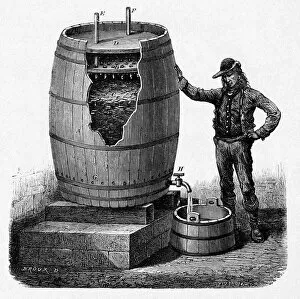
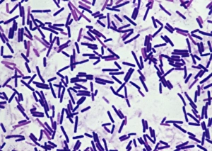
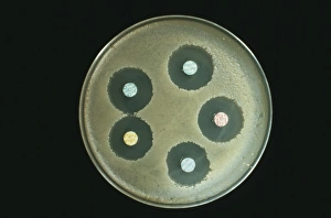
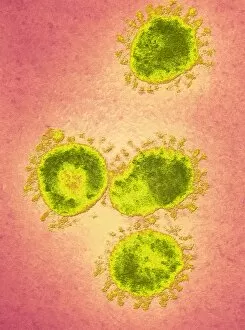
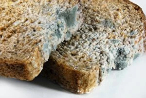
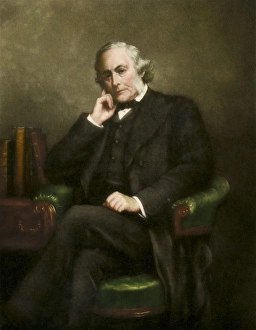
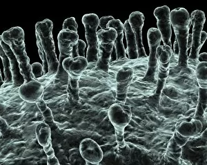
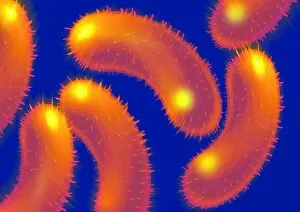
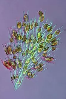
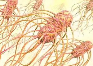
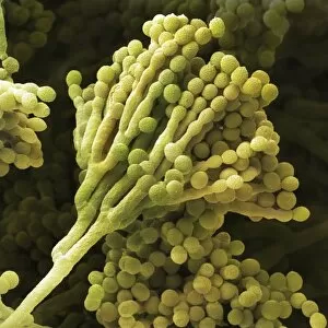
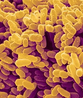
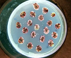
In the 19th century, microbial discoveries revolutionized the field of microbiology. From Louis Pasteur's groundbreaking research on bacterial diseases to the production of vinegar, these tiny organisms held immense significance. Microbiology caricatures from that era depicted scientists peering through microscopes, unraveling the mysteries hidden within microbial worlds. These illustrations captured their fascination and dedication towards understanding these minuscule creatures. One such breakthrough was made by Louis Pasteur himself, whose work focused on bacterial diseases. His findings paved the way for advancements in medicine and saved countless lives. The Century Illustrated Monthly Magazine featured his achievements, highlighting his contributions to science. Nature also showcased microbial wonders in Yellowstone National Park, Wyoming. Thermal bacteria mats thrived in warm waters while tourists marveled at steam rising from hot springs. This unique ecosystem demonstrated how life could flourish even under extreme conditions. The park's Black Sand Basin further exemplified this phenomenon as vibrant microbial mats grew amidst its geothermal features. These colorful displays were a testament to nature's resilience and adaptability. Microbes weren't always beneficial; some posed serious threats like anthrax and botulism bacteria. Scientists tirelessly researched antibiotics to combat these deadly infections, striving to protect public health worldwide. Even viruses didn't escape scrutiny as infectious bronchitis virus (IBV) came under intense investigation using transmission electron microscopy (TEM). This technique allowed researchers to observe viral structures with incredible detail, aiding in developing effective treatments against viral illnesses. Thermophile bacterial mats found near Grand Prismatic Spring added another layer of intrigue to Yellowstone's diverse landscape. Tourists marveled at both the beauty and scientific importance of these thriving communities living amidst scorching temperatures. From caricatures depicting early microbiologists' curiosity to modern-day research combating infectious diseases, microbes have captivated our attention throughout history. Their impact spans various fields - from medical breakthroughs to ecological wonders - reminding us that sometimes it is the smallest things that hold the greatest significance.