Fraud Blocker Skip to main content

Metacarpals Collection

"Exploring the Marvels of Metacarpals: A Comparative Study Across Mammalian Hands" In 1898

Background imageMetacarpals Collection: Comparing the bones in the hands of nine different mammals, 1898 (colour litho)

Comparing the bones in the hands of nine different mammals, 1898 (colour litho)
1065622 Comparing the bones in the hands of nine different mammals, 1898 (colour litho) by Haeckel, Ernst (1834-1919); Private Collection; (add.info.: 1.Man, 2.Gorilla, 3. Orangutang, 4. dog, 5)

Background imageMetacarpals Collection: Normal hand, digital X-ray

Normal hand, digital X-ray
Normal hand. Digital X-ray of the normal right hand of a patient in their late twenties. Various sets of data relating to the image can be seen around the edges of the X-ray

Background imageMetacarpals Collection: Ischaemia, digital angiogram

Ischaemia, digital angiogram
image d angiographie numerisee( non soustraite) de la main gauche, en vue de face, par catheterisme hyperselectif des arteres du membre superieur gauche, sous anasthesie generale

Background imageMetacarpals Collection: Bones of the human skeleton

Bones of the human skeleton
Bones of the skeleton. Artwork from Juan Valverde de Amuscos textbook Anatomia del Corpo Humano (Anatomy of the Human Body), published 1560. Fig. I is a view of the spine, from the left side. Figs

Background imageMetacarpals Collection: Illustration of right hand bones

Illustration of right hand bones
Medicine: Human anatomy, right hand bones. Drawing

Background imageMetacarpals Collection: Wrist joint anatomy, artwork C016 / 6549

Wrist joint anatomy, artwork C016 / 6549
Wrist joint anatomy. Artwork of the bones (yellow) and ligaments (blue) of the wrist joint, in longitudinal section. The left hand has been sectioned in a palmar aspect to expose the synovial

Background imageMetacarpals Collection: Hand ligaments and tendons, artwork

Hand ligaments and tendons, artwork
Hand bones and cartilage. Artwork of the bones and cartilage (white, ends of bones) of the hand and the wrist joint from the rear (dorsal) side

Background imageMetacarpals Collection: Hand anatomy, illustration C018 / 0796

Hand anatomy, illustration C018 / 0796
Hand anatomy. Illustration showing the bones, nerves and blood vessels of the hand. There are 27 bones, made up of 14 phalanges on the fingers and thumb

Background imageMetacarpals Collection: Forearm muscles, artwork

Forearm muscles, artwork
Forearm muscles. Anatomical computer artwork showing the bones (white), muscles (red), and tendons and ligaments (blue) of a human forearm, wrist and hand

Background imageMetacarpals Collection: Wrist pain, artwork C013 / 8822

Wrist pain, artwork C013 / 8822
Wrist pain, computer artwork

Background imageMetacarpals Collection: Wrist ligaments, artwork C013 / 4651

Wrist ligaments, artwork C013 / 4651
Wrist ligaments. Computer artwork of the bones of a human hand, showing the ligaments (white) holding together the bones of the wrist

Background imageMetacarpals Collection: Forearm muscle, artwork C013 / 4585

Forearm muscle, artwork C013 / 4585
Forearm muscle. Computer artwork of an anterior view of a human arm, showing the flexor pollicis longus muscle (red) of the forearm and its attachments to the bones of the arm, wrist and hand

Background imageMetacarpals Collection: Adult and child hand X-rays

Adult and child hand X-rays
Adult and child hands. Coloured X-rays of healthy human hands at 3 years (left) and at 20 years. Bones are green with flesh red

Background imageMetacarpals Collection: Thalassaemia, X-ray

Thalassaemia, X-ray
Thalassaemia. Coloured hand X-ray of a patient with thalassaemia, also known as Cooleys anaemia. The bones of the hand show demineralization (lighter patches)

Background imageMetacarpals Collection: Healthy adult hand, X-ray

Healthy adult hand, X-ray
Healthy hand. Coloured X-ray of the healthy hand of a 60 year old man, showing the skeleton of bones. The fleshy outline of the hand is seen

Background imageMetacarpals Collection: Hand bones

Hand bones. Computer artwork of a healthy hand and wrist joint. There are eight wrist (carpal) bones (bottom right). The carpal bones articulate with the bones of the forearm

Background imageMetacarpals Collection: Fractured palm bones of hand, X-ray

Fractured palm bones of hand, X-ray
Fractured palm bones. Coloured X-ray of the hand of a 35 year old man showing three fractured meta- carpal bones. The skeleton of the hand is seen

Background imageMetacarpals Collection: Osteoid osteoma, X-ray

Osteoid osteoma, X-ray
Osteoid osteoma. Coloured X-ray of an osteoid osteoma (white, centre left) in the second phalange bone of the middle finger

Background imageMetacarpals Collection: Arthritic hand

Arthritic hand, computer artwork. The finger and wrist joints are inflamed and swollen. Rheumatoid arthritis is caused by the immune system attacking the bodys own tissues

Background imageMetacarpals Collection: Arthritic hand, conceptual artwork

Arthritic hand, conceptual artwork. Arthritis is a degenerative disease that results in the loss of cartilage between joints (red), causing pain and inflammation

Background imageMetacarpals Collection: Fingertip laceration injuries, X-rays

Fingertip laceration injuries, X-rays
Fingertip laceration injuries, coloured X-rays. Both X-rays show the same hand, which suffered severe lacerations (blunt trauma cuts) to the tips of the middle fingers (green)

Background imageMetacarpals Collection: Hand anatomy, artwork

Hand anatomy, artwork
hand, bone, skeleton

Background imageMetacarpals Collection: Human skeleton, artwork

Human skeleton, artwork
Human skeleton. Computer artwork of a human skeleton, with a close-up of its hand. The skull (upper centre) and rib cage (centre and lower centre) can also be seen


All Professionally Made to Order for Quick Shipping

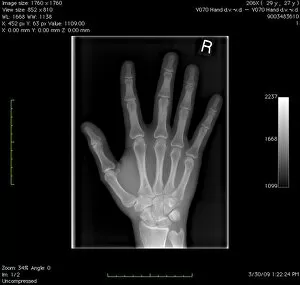
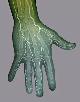
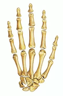
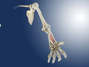
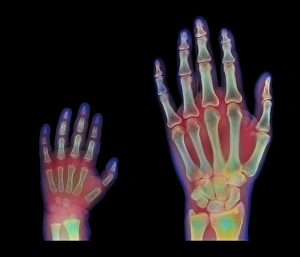
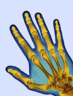
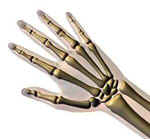
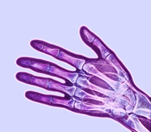
"Exploring the Marvels of Metacarpals: A Comparative Study Across Mammalian Hands" In 1898, a groundbreaking study delved into the intricate world by comparing these hand bones across nine different mammals. Through vivid color lithographs, this research shed light on the fascinating similarities and differences in their structures. Fast forward to modern times, where digital X-rays have revolutionized our understanding of metacarpals. These high-resolution images provide an unparalleled glimpse into the normal hand's internal composition, unraveling its complex network of bones. But it doesn't stop there. Digital angiograms have allowed us to explore ischaemia within metacarpals. By visualizing blood flow patterns, we gain crucial insights into potential circulatory issues affecting these vital hand bones. Let's not forget about our own human skeleton—metacarpals play a pivotal role here too. Illustrations beautifully depict the arrangement and connections between these finger-supporting bones within our hands' framework. Zooming in further, detailed illustrations focus solely on right-hand bone anatomy—a captivating exploration that showcases each metacarpal's unique characteristics and functions. The wrist joint—an essential component for metacarpal mobility—is dissected through meticulous artwork (C016 / 6549). This comprehensive analysis highlights how various ligaments contribute to stability while allowing flexible movements necessary for daily activities. Ligaments and tendons work harmoniously within our hands—artwork brings them to life visually (C018 / 0796). Understanding their roles helps us appreciate how they support metacarpals' functionality with precision and finesse. Moving beyond just studying individual elements, forearm muscles also influence metacarpi functioning. Artistic renderings capture their complexity (repeated twice), showcasing how they interact seamlessly with these finger-supporting bones during gripping motions or delicate tasks like writing or painting. Unfortunately, wrist pain can plague individuals, hindering metacarpal movements.