Fraud Blocker Skip to main content

Membranes Collection (page 2)

"Exploring the Intricate World of Membranes: From Chloroplasts to Bacterial Meningitis" Delving into the depths of cellular structures

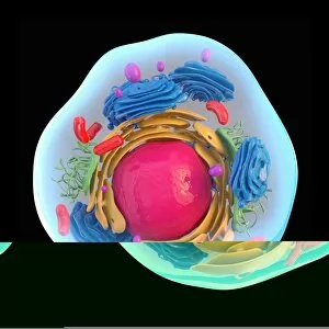
Background imageMembranes Collection: Animal cell, artwork C013 / 9985

Animal cell, artwork C013 / 9985
Animal cell. Computer artwork showing the cell organelles found inside a typical animal cell. The nucleus (large round) can be seen at centre

Background imageMembranes Collection: Animal cell, artwork C013 / 9984

Animal cell, artwork C013 / 9984
Animal cell. Computer artwork showing the cell organelles found inside a typical animal cell. The nucleus (large round) can be seen at centre

Background imageMembranes Collection: Mitochondria, artwork C013 / 4993

Mitochondria, artwork C013 / 4993
Mitochondria, computer artwork. Mitochondria are a type of organelle found in the cytoplasm of eukaryotic cells. They oxidise sugars and fats to produce energy in a process called respiration

Background imageMembranes Collection: Mitochondria, artwork C013 / 4991

Mitochondria, artwork C013 / 4991
Mitochondria, computer artwork. Mitochondria are a type of organelle found in the cytoplasm of eukaryotic cells. They oxidise sugars and fats to produce energy in a process called respiration

Background imageMembranes Collection: Retinal rod cell, TEM C013 / 4805

Retinal rod cell, TEM C013 / 4805
Retinal rod cell. Transmission electron micrograph (TEM) of a section through a rod cell from the retina of an eye, showing the inner segment (bottom) filled with mitochondria (green)

Background imageMembranes Collection: Retinal rod cell, TEM C013 / 4804

Retinal rod cell, TEM C013 / 4804
Retinal rod cell. Transmission electron micrograph (TEM) of a section through a rod cell from the retina of an eye, showing the inner segment (bottom) filled with mitochondria (green)

Background imageMembranes Collection: Ostrich (Struthio camelus) adult, close-up of head, with nictitating membrane fully closed (captive)

Ostrich (Struthio camelus) adult, close-up of head, with nictitating membrane fully closed (captive)

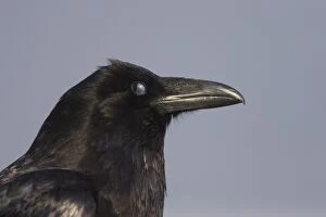
Background imageMembranes Collection: Common Raven (Corvus corax principalis) adult, close-up of head, nictitating membrane over eye

Common Raven (Corvus corax principalis) adult, close-up of head, nictitating membrane over eye, Rocky Mountains, Alberta, Canada, june

Background imageMembranes Collection: Booted Eagle (Hieraaetus pennatus) adult, close-up of eye with nictitating membrane, Spain, july

Booted Eagle (Hieraaetus pennatus) adult, close-up of eye with nictitating membrane, Spain, july

Background imageMembranes Collection: Adipose tissue, SEM

Adipose tissue, SEM
Adipose tissue. Coloured scanning electron micrograph (SEM) of adipose tissue. The tissue consists of adipocyte cells, whose lipid (fat) contents has been largely removed

Background imageMembranes Collection: Brain anatomy, 14th century artwork

Brain anatomy, 14th century artwork. The cranial vault (top of the skull, left) has been removed and two of the skull sutures (zig-zag lines) are shown

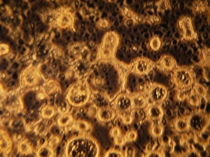
Background imageMembranes Collection: Simulated space ice

Simulated space ice. Light micrograph of vesicles (round structures) in ice formed under conditions simulating those in space

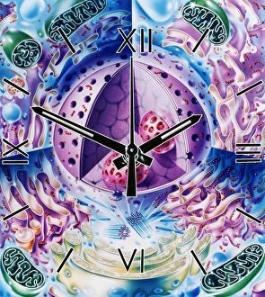
Background imageMembranes Collection: Cellular clock

Cellular clock. Conceptual artwork of a clock and the internal structures (organelles) of an animal cell. This could represent the rate of metabolic cell reactions

Background imageMembranes Collection: Cell nucleus, TEM

Cell nucleus, TEM
Cell nucleus. Coloured transmission electron micrograph (TEM) of a section through a cell, showing the nucleus (large, spherical), and mitochondria (green)

Background imageMembranes Collection: Head anatomy, artwork

Head anatomy, artwork
Head anatomy. Artwork showing various aspects of human anatomy in a female head seen from the side. The bones (white) include the lower jaw bone, the bones of the palate, the cheek bones

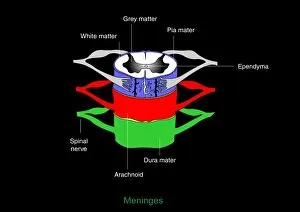
Background imageMembranes Collection: Meninges, artwork

Meninges, artwork
Meninges. Computer artwork showing the meninges surrounding the spinal cord. The meninges are the three membranes that envelop the brain and spinal cord

Background imageMembranes Collection: Immune system, artwork

Immune system, artwork
Immune system. Computer artwork showing how T-cells (right), antibodies (Y-shaped) and antigen-presenting cells (APC, left) interact during an immune response

Background imageMembranes Collection: Chloroplast structures, artwork

Chloroplast structures, artwork
Chloroplast structures. Cutaway artwork showing the internal structure of a chloroplast, the organelle in plant cells responsible for photosynthesis

Background imageMembranes Collection: Mitochondrion, artwork

Mitochondrion, artwork
Mitochondrion. Cutaway artwork showing the internal structure of a mitochondrion. This structure, found in eukaryotic cells, is the site of energy production

Background imageMembranes Collection: Animal cell processes, artwork

Animal cell processes, artwork
Animal cell processes. Cutaway artwork showing the structures inside an animal cell and four different processes that take place inside it or on its membrane (all marked by magnifying glasses)

Background imageMembranes Collection: Multiverse layers, artwork

Multiverse layers, artwork. The multiverse, also called the meta-universe or metaverse, is the hypothetical set of possible universes, and includes all of space and time

Background imageMembranes Collection: Desalination Reverse Osmosis filter

Desalination Reverse Osmosis filter
Desalination plant. A bank of Reverse Osmosis membrane filters. This facility turns salt water into drinking water using the Reverse Osmosis Process

Background imageMembranes Collection: Bacterial cell structure, artwork

Bacterial cell structure, artwork
Bacterial cell structure. Computer artwork showing the cell structure and components (organelles) of a typical rod-shaped bacteria (bacillus). Not all bacteria have a flagellum (long, tail-like)

Background imageMembranes Collection: Bacterial cell wall comparison, artwork

Bacterial cell wall comparison, artwork
Bacterial cell wall comparison. Computer artwork comparing the structure of the cell wall from a gram-negative bacterium (left) with that of a gram-positive bacterium (right)

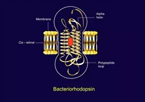
Background imageMembranes Collection: Bacteriorhodopsin, diagram

Bacteriorhodopsin, diagram
Bacteriorhodopsin. Diagram of the molecular structure of bacteriorhodopsin, a protein found in primitive micro-organisms known as Archaea

Background imageMembranes Collection: Bacteriorhodopsin, artwork

Bacteriorhodopsin, artwork
Bacteriorhodopsin. Artwork of the molecular structure of bacteriorhodopsin, a protein found in primitive micro-organisms known as Archaea

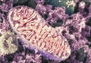
Background imageMembranes Collection: Mitochondrion, SEM

Mitochondrion, SEM
Mitochondrion. Coloured scanning electron micrograph (SEM) of a mitochondrion in a nerve cell. Mitochondria are a type of organelle found in the cytoplasm of eukaryotic cells

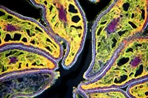
Background imageMembranes Collection: Mature poppy ovary, light micrograph

Mature poppy ovary, light micrograph
Mature poppy ovary. Light micrograph (LM) of a cross-section through a mature poppy ovary, which is incompletely divided by septa (membranes) that do not meet in the centre

Background imageMembranes Collection: Intestinal smooth muscle cells, TEM

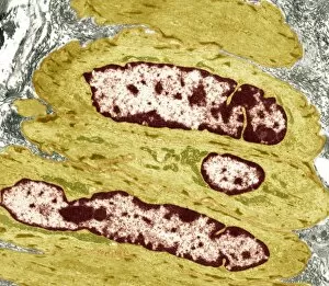
Intestinal smooth muscle cells, TEM
Intestinal smooth muscle cells. Coloured transmission electron micrograph (TEM) of a section through several smooth muscle cells from the intestines

Background imageMembranes Collection: Spinal cord, transverse section

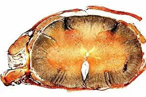
Spinal cord, transverse section
Spinal cord. Light micrograph of a transverse section through a human spinal cord. The spinal cord has a central canal around which are the dorsal and ventral horns (orange)

Background imageMembranes Collection: Born with a Caul

Born with a Caul
A child born with a caul - i.e. delivered with the membranes intact : the caul has great value in popular belief


All Professionally Made to Order for Quick Shipping

"Exploring the Intricate World of Membranes: From Chloroplasts to Bacterial Meningitis" Delving into the depths of cellular structures, we uncover the mesmerizing beauty of chloroplast membranes through intricate artwork. Shedding light on a medical mystery, an MRI scan reveals the devastating effects of bacterial meningitis on brain membranes. Peering into the microscopic realm, a TEM image showcases the delicate and vital role played by intestinal microvilli in nutrient absorption. Safeguarding our most precious organ, brain meninges shield and protect our cognitive powerhouse with unwavering dedication. Unveiling energy powerhouses within cells, SEM imaging captures stunning details of mitochondria's complex membrane structure. Witnessing an artistic representation of "Invasion de la gorge et du larynx par les fausses membranes, " we are reminded of the harrowing impact that diphtheria can have on throat and larynx membranes (colour litho). Journeying back to life's earliest stages, an engraving offers a magnified view of a four-week-old human embryo with its protective membranes opened wide. Exploring bone anatomy at its core, a cross-sectional diagram highlights femur's osteon network alongside veins and marrow while emphasizing membrane significance. Revealing nature's architectural marvels once more, SEM imagery unveils intricate details within chloroplasts' membranous structures responsible for photosynthesis. Appreciating artistry in scientific depictions, F007 / 1477 artwork portrays cell membrane lipid bilayer intricacies - showcasing their importance in cellular function. Returning to safeguard our brains yet again; let us marvel at brain meninges' resilience as they continue their tireless duty day after day. Revisiting mitochondria through SEM imaging allows us to witness their awe-inspiring complexity once more – reminding us of their crucial role in cellular energy production.