
Meiosis Collection
Meiosis, the intricate dance of genetic material separation, is beautifully depicted in these cross-section biomedical illustrations


All Professionally Made to Order for Quick Shipping
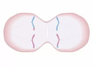
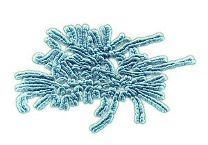
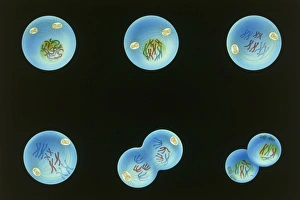
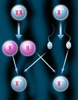
Meiosis, the intricate dance of genetic material separation, is beautifully depicted in these cross-section biomedical illustrations. In the first image, we witness the duplicated chromosomes with their unique mixture of genetic material and delicate threads forming within the cell. These threads play a crucial role as they diligently pull apart pairs of chromosomes to ensure proper distribution. Moving on to the next illustration, we observe how meiosis progresses further as the cell divides into two new cells. Remarkably, each newly formed cell carries a complete set of 23 duplicated chromosomes inherited from its parent cell. This meticulous process ensures that genetic information remains intact and distributed equally among offspring. As we delve deeper into meiosis through another captivating artwork, our attention is drawn to duplicated chromosomes lined up meticulously. Additional threads attach themselves to these duplicates, orchestrating their separation into two distinct single chromosomes. The precision exhibited here showcases nature's remarkable ability to maintain genetic stability. Intriguingly, meiosis doesn't stop at just producing two cells; it continues its symphony by dividing those cells once again. The subsequent illustration reveals four cells emerging from this division process - each containing half of the original cell's genetic material, and is through this step that diversity arises and contributes significantly to evolution's tapestry. Beyond these mesmerizing illustrations lies an Early Devonian bay teeming with thousands of Aglaophyton organisms - a testament to life's complexity and interconnectedness throughout history. These artistic renderings provide us with glimpses into various stages of meiosis: from an eight-cell embryo brimming with potential for growth and development to spermatogonia captured under high-resolution transmission electron microscopy (TEM). Each image encapsulates different facets of this fundamental biological process. Whether observed through scanning electron microscopy (SEM) or brought forth by talented artists' hands, these visual representations invite us into the world where cellular intricacies unfold during meiotic events.