
Medical Instrument Collection
In the 19th century, a female medical student delves into the world of dissection, as depicted in a captivating wood engraving


All Professionally Made to Order for Quick Shipping
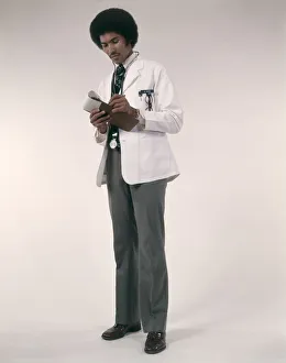
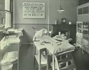
In the 19th century, a female medical student delves into the world of dissection, as depicted in a captivating wood engraving. The image showcases her determination and passion for learning about the human body. Another intriguing artwork from that era is "The Anatomist" by Thomas Rowlandson. This piece captures the intensity and precision required in anatomical studies during this time. Joseph-Claude-Anthelme Recamier's vaginal speculum, made of wood and metal, serves as a reminder of the advancements made in gynecological instruments. It symbolizes progress in women's healthcare and highlights the contributions of individuals like Recamier. Similarly, Antoine Jobert de Lamballe's vaginal speculum crafted from metal and ivory demonstrates further innovation in this field. These instruments played crucial roles in improving diagnostic accuracy and treatment options for women. An early 18th-century brass and copper apothecary's pestle and mortar reflects the importance of compounding medicines accurately. Pharmacists relied on these tools to create customized remedies tailored to patients' needs. Doctor Antommarchi utilized surgical instruments for Emperor Napoleon I's dissection, showcasing both his expertise as a physician and historical significance surrounding one of history's most prominent figures. A porcelain bleeding bowl from Strasbourg dating back to c. 1760 reminds us of traditional bloodletting practices prevalent at that time. This artifact represents an era when medical treatments were still evolving but held cultural significance nonetheless. Moving forward through history brings us to an illustration depicting an African-American man doctor wearing a white coat while writing prescriptions with his stethoscope around his neck. This powerful image challenges stereotypes while highlighting diversity within medicine. An intricate illustration featuring lymphocytes emphasizes our understanding of immune system components critical for maintaining health or combating diseases effectively. Transporting ourselves to London in 1914 reveals two operating rooms: one located at Woolwich School Treatment Centre and another at Fulham School Treatment Centre.