
Maxillae Collection
The Pictorial Museum of Animated Nature takes us on a captivating journey through the intricate world of maxillae


All Professionally Made to Order for Quick Shipping
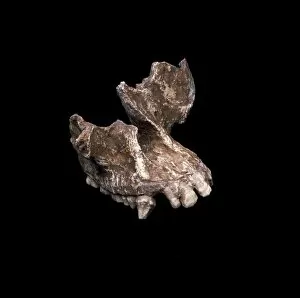
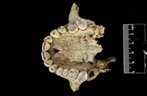
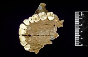
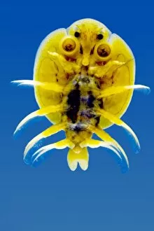
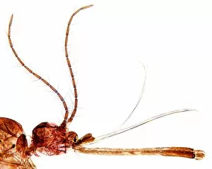
The Pictorial Museum of Animated Nature takes us on a captivating journey through the intricate world of maxillae. From the cranial fragment of Homo heidelbergensis, also known as the Rhodesian or Broken Hill Man, to the stunning artwork depicting an orangutan skull, these pages are filled with fascinating glimpses into the diversity and complexity of this vital anatomical feature. One cannot help but marvel at the Hominoid cranial fragment C016/5608 - a relic from our ancient past that offers clues about our evolutionary history. The delicate structure and unique shape of this maxilla hint at the remarkable adaptations that allowed early humans to thrive in their environments. Moving forward in time, we encounter another specimen from Homo heidelbergensis - Goughs Cave 139. This particular maxilla serves as a reminder of our shared ancestry with this enigmatic species, highlighting both our similarities and differences. But it is not only human maxillae that captivate us within these pages; nature's creations offer their own wonders. A Sivapithecus meteai cranial fragment provides insight into primate evolution, while microscopic images reveal unexpected beauty in unexpected places. A crustacean parasite caught under a microscope showcases its intricate mouthparts, while bee mouthparts demonstrate nature's ingenuity for nectar collection. The gnat head reveals astonishing detail in its tiny features, and even a cockroach's head becomes an object worthy of admiration when seen up close. In each image captured within The Pictorial Museum of Animated Nature lies a story waiting to be discovered – tales told through bones and micrographs alike. These snapshots remind us that every living creature has its place in Earth's grand tapestry, connected by threads woven through millions of years. So let us turn these pages with curiosity and wonderment as we explore the diverse world – symbols not just of anatomy but also reminders that life itself is a masterpiece, waiting to be uncovered.